

")


磁共振成像在输卵管浆液性腺癌诊断中的应用:临床病例
- 作者: Mynko O.I.1,2, Gonchar A.P.1,3, Nechaev V.A.3, Kulikova E.A.3, Yudin A.L.2,3, Yumatova E.A.2,3
-
隶属关系:
- Research and Practical Clinical Center for Diagnostics and Telemedicine Technologies
- Pirogov Russian National Research Medical University
- City Clinical Oncological Hospital 1
- 期: 卷 5, 编号 4 (2024)
- 页面: 882-892
- 栏目: 临床病例及临床病例的系列
- URL: https://bakhtiniada.ru/DD/article/view/309843
- DOI: https://doi.org/10.17816/DD628840
- ID: 309843
如何引用文章

详细
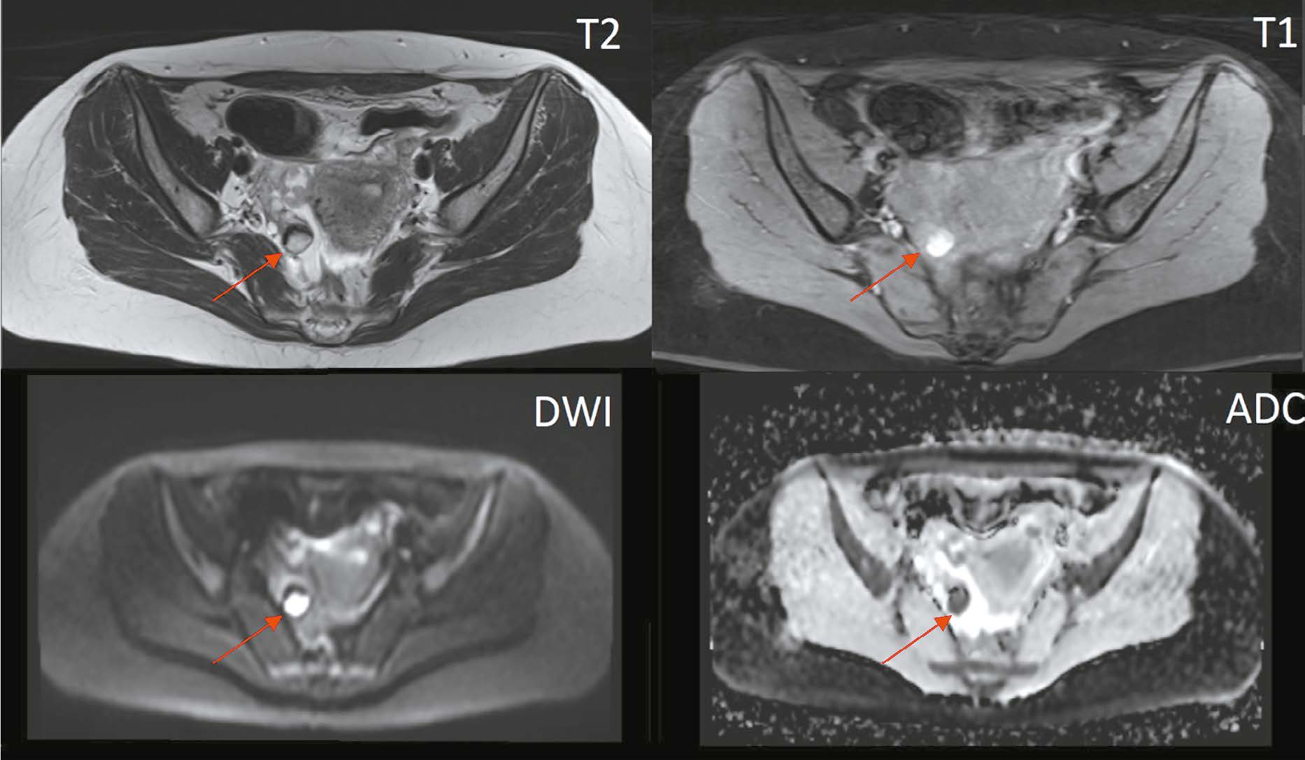
输卵管浆液性腺癌是一种极其罕见且难以诊断的女性生殖系统恶性肿瘤。这种病理通常无症状或伴有非特异性临床表现,包括浆液性血性阴道分泌物、下腹部和骨盆绞痛。这些症状在文献中被称为 “Latzko三联征»,被认为是输卵管癌的标志性症状,但只有不到15%的患者会同时出现这些症状。因其发病率低,临床症状不明显,导致大量诊断错误,或发现时疾病已处于晚期,从而大大恶化了患者的预后。只有4%的病例可在术前得到准确诊断。本临床观察描述了一例输卵管浆液性腺癌病例,该病例具有“Latzko三联征”的所有表现和磁共振成像,因此在术前阶段就被怀疑为输卵管浆液性腺癌。
作者简介
Oleg I. Mynko
Research and Practical Clinical Center for Diagnostics and Telemedicine Technologies; Pirogov Russian National Research Medical University
编辑信件的主要联系方式.
Email: o.mynko@icloud.com
ORCID iD: 0009-0005-3984-4045
SPIN 代码: 3556-3510
MD
俄罗斯联邦, Moscow; MoscowAnna P. Gonchar
Research and Practical Clinical Center for Diagnostics and Telemedicine Technologies; City Clinical Oncological Hospital 1
Email: a.gonchar@npcmr.ru
ORCID iD: 0000-0001-5161-6540
SPIN 代码: 3513-9531
MD
俄罗斯联邦, Moscow; MoscowValentin A. Nechaev
City Clinical Oncological Hospital 1
Email: dfkz2005@gmail.com
ORCID iD: 0000-0002-6716-5593
SPIN 代码: 2527-0130
MD, Cand. Sci. (Medicine), Head of complex diagnostic center
俄罗斯联邦, MoscowEvgeniya A. Kulikova
City Clinical Oncological Hospital 1
Email: kulikovaEA14@zdrav.mos.ru
ORCID iD: 0000-0002-0319-4934
SPIN 代码: 2884-4803
MD
俄罗斯联邦, MoscowAndrey L. Yudin
Pirogov Russian National Research Medical University; City Clinical Oncological Hospital 1
Email: prof_yudin@mail.ru
ORCID iD: 0000-0002-0310-0889
SPIN 代码: 6184-8284
MD, Dr. Sci. (Medicine), Professor
俄罗斯联邦, Moscow; MoscowElena A. Yumatova
Pirogov Russian National Research Medical University; City Clinical Oncological Hospital 1
Email: yumatova_ea@mail.ru
ORCID iD: 0000-0002-6020-9434
SPIN 代码: 8447-8748
MD, Cand. Sci. (Medicine)
俄罗斯联邦, Moscow; Moscow参考
- Meinhold Heerlein I, Fotopoulou C, Harter P, et al. The new WHO classification of ovarian, fallopian tube, and primary peritoneal cancer and its clinical implications. Arch Gynecol Obstet. 2016;293(4):695–700. doi: 10.1007/s00404-016-4035-8
- Burghardt E, Girardi F, Lahousen M, et al. Patterns of pelvic and paraaortic lymph node involvement in ovarian cancer. Gynecol Oncol. 1991;40(2):103–106. doi: 10.1016/0090-8258(91)90099-q
- Stasenko M, Fillipova O, Tew W. Fallopian Tube Carcinoma. J Oncol Pract. 2019;15(7):375–382. doi: 10.1200/JOP.18.00662
- Reid B, Permuth J, Sellers T. Epidemiology of ovarian cancer: a review. Cancer Biol Med. 2017;14(1):9–32. doi: 10.20892/j.issn.2095-3941.2016.0084
- Ulrikh EA, Papunidi МD, Urmancheeva AF, Matsko DE. Fallopian tube carcinoma: clinical and morphological features, analysis of 69 cases. Voprosy onkologii. 2014;60(3):375–378. EDN: SJTCOH
- Kim MY, Rha SE, Oh SN, et al. MR Imaging findings of hydrosalpinx: a comprehensive review. Radiographics. 2009;29(2):495–507. doi: 10.1148/rg.292085070
- Riska A, Leminen A. Updating on primary fallopian tube carcinoma. Acta Obstet Gynecol Scand. 2007;86:1419–1426. doi: 10.1080/00016340701771034
- PDQ Adult Treatment Editorial Board. Ovarian Epithelial, Fallopian Tube, and Primary Peritoneal Cancer Treatment (PDQ®): Health Professional Version. 2023. In: PDQ Cancer Information Summaries [Internet]. Bethesda (MD): National Cancer Institute (US), 2002. Available from: https://www.ncbi.nlm.nih.gov/books/NBK66007/
- Kalampokas E, Kalampokas T, Tourountous I. Primary fallopian tube carcinoma. Eur J Obstet Gynecol Reprod Biol. 2013;169(2):155–161. doi: 10.1016/j.ejogrb.2013.03.023
- Carvalho J, Moretti Marques R, Filho A. Adnexal mass: diagnosis and management. Rev Bras Ginecol Obstet. 2020;42(7):438–443. doi: 10.1055/s-0040-1715547
- Anthoulakis C, Nikoloudis N. Pelvic MRI as the ”gold standard” in the subsequent evaluation of ultrasound indeterminate adnexal lesions: a systematic review. Gynecol Oncol. 2014;132(3):661–668. doi: 10.1016/j.ygyno.2013.10.022
- Nishino M, Hayakawa K, Minami M, et al. Primary retroperitoneal neoplasms: CT and MR imaging findings with anatomic and pathologic diagnostic clues. Radiographics. 2003;23(1):45–57. doi: 10.1148/rg.231025037
- Sadowski E, Thomassin Naggara I, Rockall A, et al. O-RADS MRI Risk Stratification System: Guide for Assessing Adnexal Lesions from the ACR O-RADS Committee. Radiology. 2022;303(1):35–47. doi: 10.1148/radiol.204371
- Bulanov MN, Chekalova MA, Mazurkevich MN, Vetsheva NN. Primenenie sistemy O-RADS pri ultrazvukovom issledovanii pridatkov matki. Moscow: Research and Practical Clinical Center for Diagnostics and Telemedicine Technologies of the Moscow Health Care Department, 2022. 27 p. (In Russ.) EDN: BUBNGP
- Veloso G, Dias F, Lucas R, Cunha T. Primary fallopian tube carcinoma: review of MR imaging findings. Insights Imaging. 2015;6(4):431–439. doi: 10.1007/s13244-015-0416-y
- Duska LR, Kohn EC. The new classifications of ovarian, fallopian tube, and primary peritoneal cancer and their clinical implications. Ann Oncol. 2017;28(suppl_8):viii8-viii12. doi: 10.1093/annonc/mdx445
- Nudnov NV, Ivashina SV, Aksenova SP. Radiation methods in the diagnosis of primary and recurrent malignant ovarian struma: A case report. Digital Diagnostics. 2023;4(2):214−225. EDN: YNASOM doi: 10.17816/DD322846
- Klinicheskie rekomendatsii MZ RF "Rak yaichnikov / rak matochnoi truby / pervichnyi rak bryushiny". 2022. Ministerstvo zdravookhraneniya RF. Available from: https://oncology.ru/specialist/treatment/references/actual/547.pdf
- Singh N, Gilks C, Wilkinson N, et al. Assignment of primary site in high grade serous tubal, ovarian and peritoneal carcinoma: a proposal. Histopathology. 2014;65(2):149–154. doi: 10.1111/his.12419
- Zhordania KI, Payanidi YuG, Kalinicheva EV. Two ways of the development of serous epithelial ”Ovarian” cancer. Oncogynecology. 2014;(3):42–48. EDN: TAOOQL
- Zhordania KI, Payanidi YuG, Kalinicheva EV. Novaya paradigma v etiologii seroznogo raka yaichnikov. Rossiiskii bioterapevticheskii zhurnal. (In Russ.) 2014;13(2):95–102. EDN: SNANEL
- Zhordania KI. Serous ovarian carcinoma or serous carcinoma of uterine (fallopian) tube? Oncogynecology. 2012;(3):4–9. EDN: SZRFTZ
- SEER*Explorer: An interactive website for SEER cancer statistics [Internet]. Surveillance Research Program, National Cancer Institute; 2023. Available from: https://seer.cancer.gov/statistics network/explorer/
- Tokunaga H, Mikami M, Nagase S, et al. The 2020 Japan Society of Gynecologic Oncology guidelines for the treatment of ovarian cancer, fallopian tube cancer, and primary peritoneal cancer. J Gynecol Oncol. 2021;32(2):e49. doi: 10.3802/jgo.2021.32.e49
- Kuroki L, Guntupalli SR. Treatment of epithelial ovarian cancer. BMJ. 2020;371:m3773. doi: 10.1136/bmj.m3773
- Trabert B, Coburn SB, Mariani A, et al. Reported Incidence and Survival of Fallopian Tube Carcinomas: A Population Based Analysis From the North American Association of Central Cancer Registries. J Natl Cancer Inst. 2018;110(7):750–757. doi: 10.1093/jnci/djx263
- Morozov SP, Lindenbraten LD, Gabai PG, et al. Osnovy menedzhmenta meditsinskoi vizualizatsii. Moscow: GEOTAR-Media, 2020. 432 p. EDN: ZRGBGE doi: 10.33029/9704-5247-9-MEN-2020-1-424
- Svidetelstvo o gosudarstvennoi registratsii programmy dlya EVM № 2024618494. Rossiiskaya Federatsiya. Platforma testirovaniya i obucheniya vrachei: № 2024617367. Vasilev YuA, Shulkin IM, Arzamasov KM, et al. Nauchno prakticheskii klinicheskii tsentr diagnostiki i telemeditsinskikh tekhnologii. (In Russ.) EDN: POELJA
补充文件


