

")


Machine learning techniques for breast cancer diagnosis
- Authors: Dyomin K.S.1, Germashev I.V.1
-
Affiliations:
- Volgograd State University
- Issue: Vol 5, No 3 (2024)
- Pages: 578-591
- Section: Reviews
- URL: https://bakhtiniada.ru/DD/article/view/310039
- DOI: https://doi.org/10.17816/DD625866
- ID: 310039
Cite item
Full Text

Abstract
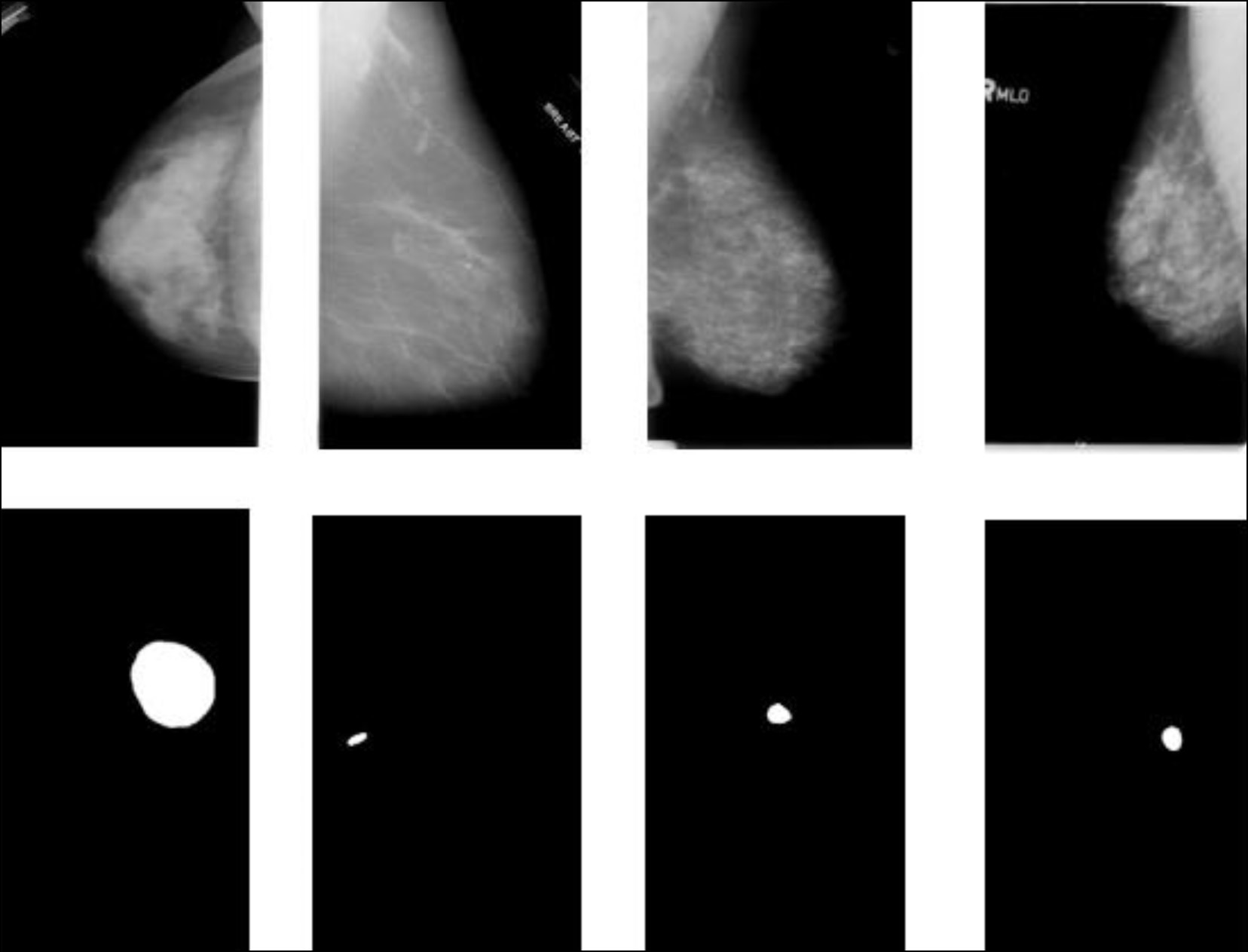
In the last few years, machine learning techniques have been attracting even greater attention in the field of diagnostics, particularly when detecting breast cancer. Relevant studies dedicated to machine learning techniques in breast cancer diagnosis were analyzed in three areas: solving secondary problems that occur in modern-day breast cancer diagnostics, role in an intelligent assessment of the patient’s condition for preliminary diagnostic decisions, and capability to detect breast cancer risk factors. The results revealed that machine learning techniques applied in breast cancer diagnosis have great potential for improving diagnostic accuracy and efficiency and solving secondary problems. The medical literature analysis has determined the parameters that are used as input data in machine learning techniques. Furthermore, the collected information will be applied to create a parameter system for breast cancer diagnosis using machine learning techniques.
Full Text
##article.viewOnOriginalSite##About the authors
Kirill S. Dyomin
Volgograd State University
Author for correspondence.
Email: diominkirill@yandex.ru
ORCID iD: 0009-0002-4571-3437
Russian Federation, Volgograd
Ilya V. Germashev
Volgograd State University
Email: germashev@volsu.ru
ORCID iD: 0000-0001-5507-8508
SPIN-code: 2489-2628
Dr. Sci. (Engineering)
Russian Federation, VolgogradReferences
- Kaprin AD, Starinsky VV, Shakhzadova AO, editors. Sostoyanie onkologicheskoj pomoshchi naseleniyu Rossii v 2022 godu. Moscow: P.A. Herzen Institute of Medical Sciences − branch of the Federal State Budgetary Institution “NMIC of Radiology” of the Ministry of Health of the Russian Federation; 2023. (In Russ.)
- Zou S, Lin Y, Yu X, et al. Genetic and lifestyle factors for breast cancer risk assessment in Southeast China. Cancer Medicine. 2023;12(14):15504–15514. doi: 10.1002/cam4.6198
- Raza SK, Sarwar SS, Syed SM, Khan NA. Classification and Segmentation of Breast Tumor Using Mask R- CNN on Mammograms. Research Square. 2021. doi: 10.21203/rs.3.rs-523546/v1
- Khan AA, Arora AS. Thermography as an Economical Alternative Modality to Mammography for Early Detection of Breast Cancer. Journal of Healthcare Engineering. 2021:5543101. doi: 10.1155/2021/5543101
- Ko C, Toh C, Brody JP. Genetic risk scores for breast cancer based on machine learning analysis of chromosomal-scale length variation. Clinical Cancer Research. 2021;27 Suppl. 5:PR-09. doi: 10.1158/1557-3265.ADI21-PR-09
- Yagin B, Yagin FH, Colak C, et al. Cancer Metastasis Prediction and Genomic Biomarker Identification through Machine Learning and eXplainable Artificial Intelligence in Breast Cancer Research. Diagnostics (Basel). 2023;13(21):3314. doi: 10.3390/diagnostics13213314
- O’Leary TJ, Mikel UV, Becker RL. Computer-assisted image interpretation: use of a neural network to differentiate tubular carcinoma from sclerosing adenosis. Modern Pathology. 1992;5(4):402–405.
- Tsochatzidis L, Costaridou L, Pratikakis I. Deep Learning for Breast Cancer Diagnosis from Mammograms – A Comparative Study. Journal of Imaging. 2019;5(3):37. doi: 10.3390/jimaging5030037
- Hamad YA, Simonov K, Naeem MB. Breast Cancer Detection and Classification Using Artificial Neural Networks. In: 1st Annual International Conference on Information and Sciences (AiCIS); Nov 20–21, 2018; Fallujah. P. 51–57. doi: 10.1109/aicis.2018.00022
- Ruchai AN, Kober VI, Dorofeev KA, et al. Classification of breast pathologies using a deep convolutional neural network and transfer learning. Information processes. 2020;20(4):357–365.
- Sasov DA, Zubkov AV, Orlova YuA, Tupitsyna AV. Classification of breast cancer using convolutional neural networks. Inženernyj vestnik Dona. 2023;6:730–741. (In Russ.)
- Computational Intelligence and Neuroscience. Retracted: Value of Artificial Neural Network Ultrasound in Improving Breast Cancer Diagnosis. Computational Intelligence and Neuroscience. 2023:9872174. doi: 10.1155/2023/9872174
- Zhang L, Jia Z, Leng X, Ma F. Artificial Intelligence Algorithm-Based Ultrasound Image Segmentation Technology in the Diagnosis of Breast Cancer Axillary Lymph Node Metastasis. Journal of Healthcare Engineering. 2021:8830260. doi: 10.1155/2021/8830260
- Zheng X, Yao Z, Huang Y, et al. Deep learning radiomics can predict axillary lymph node status in early-stage breast cancer. Nature Communications. 2020;11(1):1236. doi: 10.1038/s41467-020-15027-z
- Mustafin CK. Sovremennaya diagnostika zabolevanij molochnyh zhelez. Glavnyj vrač Ûga Rossii. 2014;(2):20–23. (In Russ.)
- Jochelson MS, Dershaw DD, Sung JS, et al. Bilateral contrast-enhanced dual-energy digital mammography: feasibility and comparison with conventional digital mammography and MR imaging in women with known breast carcinoma. Radiology. 2013;266(3):743–751. doi: 10.1148/radiol.12121084
- Witowski J, Heacock L, Reig B, et al. Improving breast cancer diagnostics with deep learning for MRI. Science Translational Medicine. 2022;14(664):eabo4802. doi: 10.1126/scitranslmed.abo4802
- Yurttakal AH, Erbay H, İkizceli T, Karacavus S. Detection of breast cancer via deep convolution neural networks using MRI images. Multimedia Tools and Applications. 2020;79:15555–15573. doi: 10.1007/s11042-019-7479-6
- Gourav Modanwal, Adithya Vellal, Maciej A. Mazurowski, Normalization of breast MRIs using cycle-consistent generative adversarial networks // Computer Methods and Programs in Biomedicine, Vol. 208, 2021. doi: 10.1016/j.cmpb.2021.106225
- Ceny na MRT. In: Like Doctor [Internet] [cited 2024 Jan 9]. Available from: https://like.doctor/ceny/diagnostika/mrt (In Russ).
- Diagnosticheskij mikrovolnovyj radiotermometr RTM-01-RES. In: Mikrovolnovaya radiotermometriya v medicine [Internet] [cited 2024 Jan 9]. Available from: http://www.radiometry.ru/radiometry/mammology/ (In Russ).
- Polyakov MV, Popov IE, Losev AG, Khoperskov AV. Application of computer simulation results and machine learning in analysis of microwave radiothermometry data. Mathematical Physics and Computer Simulation. 2021;24(2):27–37. doi: 10.15688/mpcm.jvolsu.2021.2.3
- Germashev IV, Dubovskaya VI, Losev AG, Popov IE. Factor analysis of the effect of signs on the accuracy of breast cancer diagnosis according to microwave radiothermometry. Caspian Journal: Management and High Technologies. 2022;(1):139–148.
- Losev AG, Medvedev DA. The use of neural networks in the diagnosis of breast cancer according to microwave radiothermometry. Modern Science and Innovation. 2019;(4):22–28.
- Shusharin AG, Morozov VV, Polovinka MP. Medical thermal imaging – modern possibilities of the method. Modern problems of science and education. 2011:(4).
- Titskaya AA, Chernov VI, Sinilkin IG, et al. Standartizirovannye metodiki radionuklidnoj diagnostiki. Mammoscintigrafiya. Moscow: NTC Amplitude; 2014. (In Russ.)
- Li J, Galazis C, Popov L, et al. Dynamic Weight Agnostic Neural Networks and Medical Microwave Radiometry (MWR) for Breast Cancer Diagnostics. Diagnostics (Basel). 2022;12(9):2037. doi: 10.3390/diagnostics12092037
- Glazunov VA. Testing the algorithm of tumor localization in breast cancer based on the results of modeling temperature fields. // XXV Regional Conference of Young Researchers of the Volgograd region : Theses of reports, Volgograd, November 20 – 13, 2020 / Editorial board: A.E. Kalinina (ed.) [et al.]. Volgograd: Volgograd State University, 2021. P:343–347. (In Russ.) EDN: DJQDLK
- Zamechnik TV, Losev AG, Levshinsky VV. Results of optimization of diagnostic signs of breast cancer detected by microwave radiothermometry. Medical News of North Caucasus. 2019;14(1.1):48–52. doi: 10.14300/mnnc.2019.14047
- Kakileti ST, Madhu HJ, Manjunath G, et al, Personalized risk prediction for breast cancer pre-screening using artificial intelligence and thermal radiomics. Artificial Intelligence in Medicine. 2020;105:101854. doi: 10.1016/j.artmed.2020.101854
- Mambou SJ, Maresova P, Krejcar O, et al. Breast Cancer Detection Using Infrared Thermal Imaging and a Deep Learning Model. Sensors (Basel). 2018;18(9):2799. doi: 10.3390/s18092799
- Makarova MV, Unitsina AV. Thermal imaging of mammary glands in the assessment of volumetric formations. Vestnik of Northern (Arctic) Federal University. Series “Humanitarian and Social Sciences”. 2013;(4):44–50.
- Hussein NAK, Al-Sarray B. Deep Learning and Machine Learning via a Genetic Algorithm to Classify Breast Cancer DNA Data. Iraqi Journal of Science. 2022;63(7):3153–3168. doi: 10.24996/ijs.2022.63.7.36
- Magna AAR, Allende-Cid H, Taramasco C, et al. Application of Machine Learning and Word Embeddings in the Classification of Cancer Diagnosis Using Patient Anamnesis. IEEE Access. 2020;8:106198–106213. doi: 10.1109/ACCESS.2020.3000075
- Mortazavi SAR, Tahmasebi S, Par-Saei H, Taleie A. Machine Learning Models for Predicting Breast Cancer Risk in Women Exposed to Blue Light from Digital Screens. Journal of Biomedical Physics and Engineering. 2022;12(6):637–644. doi: 10.31661/jbpe.v0i0.2105-1341
- Mortazavi SAR. The Association of Screen Time and Female Breast Cancer – A Retrospective Case-Control Study [dissertation]. Shiraz: Shiraz University of Medical Sciences; 2021.
- Afrash MR, Bayani A, Shanbehzadeh M, et al. Developing the breast cancer risk prediction system using hybrid machine learning algorithms. Journal of Education and Health Promotion. 2022;11(1):272. doi: 10.4103/jehp.jehp_42_22
Supplementary files


