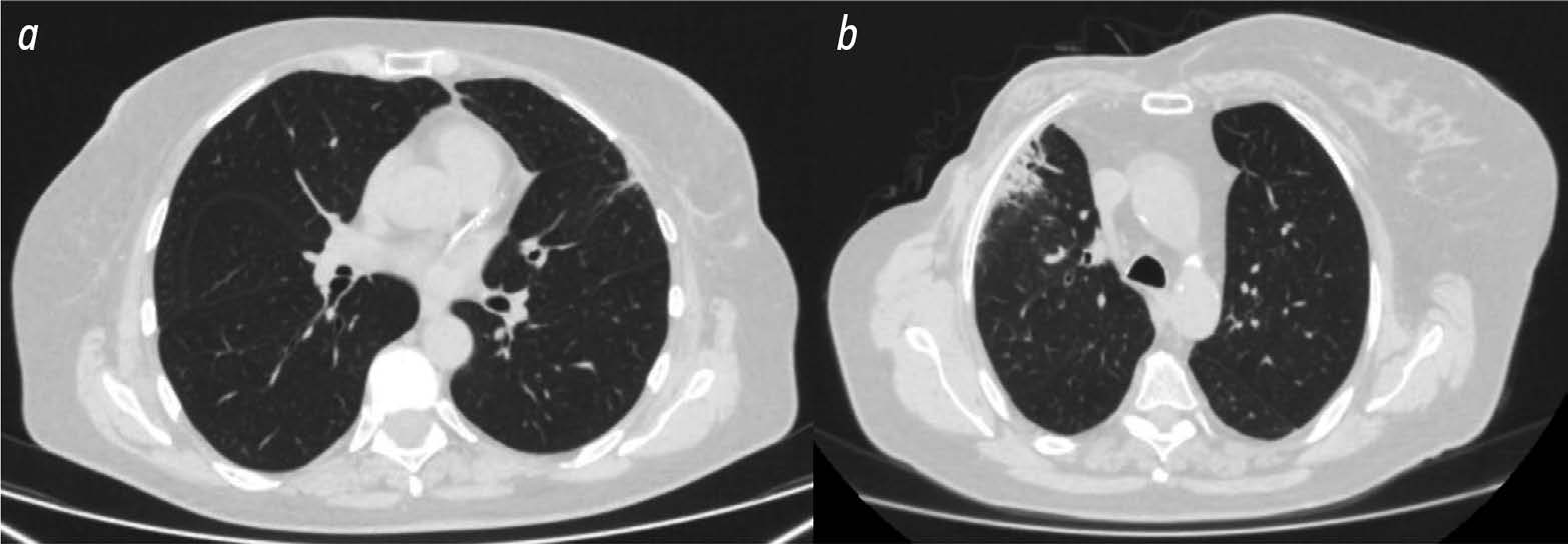
Use of radiomics and dosiomics to identify predictors of radiation induced lung injury
- Authors: Nudnov N.V.1,2,3, Sotnikov V.M.1, Ivannikov M.E.1, Shakhvalieva E.S.1, Borisov A.A.1, Ledenev V.V.4, Smyslov A.Y.1, Ananina A.V.1
-
Affiliations:
- Russian Scientific Center of Roentgenoradiology
- Russian Medical Academy of Continuous Professional Education
- Peoples’ Friendship University of Russia
- Central Clinical Military Hospital
- Issue: Vol 5, No 4 (2024)
- Pages: 752-764
- Section: Original Study Articles
- URL: https://bakhtiniada.ru/DD/article/view/309834
- DOI: https://doi.org/10.17816/DD629352
- ID: 309834
Cite item
Abstract
BACKGROUND: Radiomics is a machine learning based technology that extracts, analyzes, and interprets quantitative features from digital medical images. In recent years, dosiomics has become an increasingly common term in the literature to describe a new radiomics method. Dosiomics is a texture analysis method for evaluating radiotherapy dose distribution patterns. Most of the published research in dosiomics evaluates its use in predicting radiation induced lung injury.
AIM: The aim of the study was to identify predictors (biomarkers) of radiation induced lung injury using texture analysis of computed tomography (CT) images of lungs and chest soft tissues using radiomics and dosiomics.
MATERIALS AND METHODS: The study used data from 36 women with breast cancer who received postoperative conformal radiation therapy. Retrospectively, the patients were divided into two groups according to the severity of post radiation lung lesions. 3D Slicer was used to evaluate CT results of all patients obtained during radiation treatment planning and radiation dose distribution patterns. The software was able to unload radiomic and dosiomic features from regions of interest. The regions of interest included chest soft tissue and lung areas on the irradiated side where the dose burden exceeded 3 and 10 Gy.
RESULTS: The first group included 13 patients with minimal radiation induced lung lesions, and the second group included 23 patients with post radiation pneumofibrosis. In the lung area on the side irradiated with more than 3 Gy, statistically significant differences between the patient groups were obtained for three radiomic features and one dosiomic feature. In the lung area on the side irradiated with more than 10 Gy, statistically significant differences were obtained for 12 radiomic features and 1 dosiomic feature. In the area of chest soft tissues on the irradiated side, significant differences were obtained for 18 radiomic features and 4 dosiomic features.
CONCLUSIONS: As a result, a number of radiomic and dosiomic features were identified which were statistically different in patients with minimal lesions and pulmonary pneumofibrosis following radiation therapy for breast cancer. Based on texture analysis, predictors (biomarkers) were identified to predict post radiation lung injury and identify higher risk patients.
Full Text
##article.viewOnOriginalSite##About the authors
Nikolay V. Nudnov
Russian Scientific Center of Roentgenoradiology; Russian Medical Academy of Continuous Professional Education; Peoples’ Friendship University of Russia
Author for correspondence.
Email: nudnov@rncrr.ru
ORCID iD: 0000-0001-5994-0468
SPIN-code: 3018-2527
MD, Dr. Sci. (Medicine), Professor
Russian Federation, Moscow; Moscow; MoscowVladimir M. Sotnikov
Russian Scientific Center of Roentgenoradiology
Email: vmsotnikov@mail.ru
ORCID iD: 0000-0003-0498-314X
SPIN-code: 3845-0154
MD, Dr. Sci. (Medicine), Professor
Russian Federation, MoscowMikhail E. Ivannikov
Russian Scientific Center of Roentgenoradiology
Email: ivannikovmichail@gmail.com
ORCID iD: 0009-0007-0407-0953
SPIN-code: 3419-2977
MD
Russian Federation, MoscowElina S.-A. Shakhvalieva
Russian Scientific Center of Roentgenoradiology
Email: shelina9558@gmail.com
ORCID iD: 0009-0000-7535-8523
MD
Russian Federation, MoscowAleksandr A. Borisov
Russian Scientific Center of Roentgenoradiology
Email: aleksandrborisov10650@gmail.com
ORCID iD: 0000-0003-4036-5883
SPIN-code: 4294-4736
MD
Russian Federation, MoscowVasiliy V. Ledenev
Central Clinical Military Hospital
Email: Ledenevvv007@gmail.com
ORCID iD: 0000-0002-2856-2107
SPIN-code: 2791-0329
MD, Cand. Sci. (Medicine)
Russian Federation, MoscowAleksei Yu. Smyslov
Russian Scientific Center of Roentgenoradiology
Email: smyslov.ay@gmail.com
ORCID iD: 0000-0002-6409-6756
SPIN-code: 9341-0037
Cand. Sci. (Engineering)
Russian Federation, MoscowAlina V. Ananina
Russian Scientific Center of Roentgenoradiology
Email: vastruhina.a.v@yandex.ru
ORCID iD: 0009-0002-4562-9729
SPIN-code: 9699-7690
Russian Federation, Moscow
References
- Khmelevsky EV, Kaprin AD. The state of a radiotherapy service in Russia: Comparative analysis and prospects for development. P.A. Herzen Journal of Oncology. 2017;6(4):38-41. EDN: ZFCHGJ doi: 10.17116/onkolog20176438-41
- Kuipers ME, van Doorn Wink KCJ, Hiemstra PS, Slats AM. Predicting radiation induced lung injury in lung cancer patients — challenges and opportunities: Predicting radiation induced lung injury. Int J Radiat Oncol Biol Phys. 2023;118(3):639–649. doi: 10.1016/j.ijrobp.2023.10.044
- Mayerhoefer ME, Materka A, Langs G, et al. Introduction to Radiomics. J Nucl Med. 2020;61(4):488–495. doi: 10.2967/jnumed.118.222893
- Radiomic Features: pyradiomics v3.0.1.post15+g2791e23 documentation [Internet]. [cited 25 Nov 2023]. Available from: https://pyradiomics.readthedocs.io/en/latest/features.html#.
- Avanzo M, Stancanello J, Pirrone G, Sartor G. Radiomics and deep learning in lung cancer. Strahlenther Onkol. 2020;196(10):879–887. doi: 10.1007/s00066-020-01625-9
- Gabryś HS, Buettner F, Sterzing F, et al. Design and selection of machine learning methods using radiomics and dosiomics for normal tissue complication probability modeling of xerostomia. Front Oncol. 2018;8:35. doi: 10.3389/fonc.2018.00035
- Solodkiy VA, Nudnov NV, Ivannikov ME, et al. Dosiomics in the analysis of medical images and prospects for its use in clinical practice. Digital Diagnostics. 2023;4(3):340–355. EDN: EQRWGJ doi: 10.17816/DD420053
- Arroyo Hernández M, Maldonado F, Lozano Ruiz F, et al. Radiation induced lung injury: Current evidence. BMC Pulm Med. 2021;21(1):9. doi: 10.1186/s12890-020-01376-4
- Rahi MS, Parekh J, Pednekar P, et al. Radiation Induced Lung Injury — Current Perspectives and Management. Clin Pract. 2021;11(3):410–429. doi: 10.3390/clinpract11030056
- Yan Y, Fu J, Kowalchuk RO, et al. Exploration of radiation induced lung injury, from mechanism to treatment: a narrative review. Transl Lung Cancer Res. 2022;11(2):307–322. doi: 10.21037/tlcr-22-108
- Gladilina IA, Shabanov MA, Kravets OA, et al. Radiation Induced Lung Injury. Journal of oncology: diagnostic radiology and radiotherapy. 2020;3(2):9–18. EDN: SKOAAY doi: 10.37174/2587-7593-2020-3-2-9-18
- Nudnov NV, Sotnikov VM, Ledenev VV, Baryshnikova DV. Features a Qualitative Assessment of Radiation Induced Lung Damage by CT. Medical Visualization. 2016;(1):39–46. EDN: VWOIIB
- Ledenev VV. Methodology for quantitative assessment of radiation damage to lungs in cancer patients using CT [dissertation]. Moscow, 2023. Available from: https://www.rncrr.ru/nauka/dissertatsionnyysovet/obyavleniyaozashchitakh/upload%202023/Леденев_ Диссертация.pdf (In Russ.) EDN: YBWROM
- Zhou C, Yu J. Chinese expert consensus on diagnosis and treatment of radiation pneumonitis. Prec Radiat Oncol. 2022;6(3):262–271. doi: 10.1002/pro6.1169
- Konkol M, Śniatała P, Milecki P. Radiation induced lung injury — what do we know in the era of modern radiotherapy? Rep Pract Oncol Radiother. 2022;27(3):552–565. doi: 10.5603/RPOR.a2022.0046
- Shaymuratov RI. Radiation induced lung injury. A review. The Bulletin of Contemporary Clinical Medicine. 2020;13(3):63–73. EDN: BIZZHU doi: 10.20969/VSKM.2020.13(3).63-73
- D Slicer image computing platform [Internet]. [cited 25 Nov 2023]. Available from: https://www.slicer.org/
- Wang L, Gao Z, Li C, et al. Computed Tomography Based Delta Radiomics Analysis for Discriminating Radiation Pneumonitis in Patients With Esophageal Cancer After Radiation Therapy. Int J Radiat Oncol Biol Phys. 2021;111(2):443–455. doi: 10.1016/j.ijrobp.2021.04.047
- Begosh Mayne D, Kumar SS, Toffel S, et al. The dose response characteristics of four NTCP models: using a novel CT-based radiomic method to quantify radiation-induced lung density changes. Sci Rep. 2020;10(1):10559. doi: 10.1038/s41598-020-67499-0
- Korpela E, Liu SK. Endothelial perturbations and therapeutic strategies in normal tissue radiation damage. Radiat Oncol. 2014;9:266. doi: 10.1186/s13014-014-0266-7
- Zhao L, Sheldon K, Chen M, et al. The predictive role of plasma TGF-beta1 during radiation therapy for radiation induced lung toxicity deserves further study in patients with non small cell lung cancer. Lung Cancer. 2008;59(2):232–239. doi: 10.1016/j.lungcan.2007.08.010
- Chen S, Zhou S, Zhang J, et al. A neural network model to predict lung radiation induced pneumonitis. Med Phys. 2007;34(9):3420–3427. doi: 10.1118/1.2759601
- Jain V, Berman AT. Radiation Pneumonitis: Old Problem, New Tricks. Cancers (Basel). 2018;10(7):222. doi: 10.3390/cancers10070222
- Huang Y, Feng A, Lin Y, et al. Radiation pneumonitis prediction after stereotactic body radiation therapy based on 3D dose distribution: Dosiomics and/or deep learning-based radiomics features. Radiat Oncol. 2022;17(1):188. doi: 10.1186/s13014-022-02154-8
- Zhang Z, Wang Z, Yan M, et al. Radiomics and dosiomics signature from whole lung predicts radiation pneumonitis: A model development study with prospective external validation and decision curve analysis. Int J Radiat Oncol Biol Phys. 2023;115(3):746–758. doi: 10.1016/j.ijrobp.2022.08.047
- Puttanawarut C, Sirirutbunkajorn N, Khachonkham S, et al. Biological dosiomic features for the prediction of radiation pneumonitis in esophageal cancer patients. Radiat Oncol. 2021;16(1):220. doi: 10.1186/s13014-021-01950-y
- Liang B, Yan H, Tian Y, et al. Dosiomics: Extracting 3D spatial features from dose distribution to predict incidence of radiation pneumonitis. Front Oncol. 2019;(9):269.doi: 10.3389/fonc.2019.00269
- Liang B, Tian Y, Chen X, et al. Prediction of radiation pneumonitis with dose distribution: A convolutional neural network (CNN) based model. Front Oncol. 2020;9:1500. doi: 10.3389/fonc.2019.01500
- Adachi T, Nakamura M, Shintani T, et al. Multi institutional dose segmented dosiomic analysis for predicting radiation pneumonitis after lung stereotactic body radiation therapy. Med Phys. 2021;48(4):1781–1791. doi: 10.1002/mp.14769
- Zheng X, Guo W, Wang Y, et al. Multi omics to predict acute radiation esophagitis in patients with lung cancer treated with intensity modulated radiation therapy. Eur J Med Res. 2023;28(1):126. doi: 10.1186/s40001-023-01041-6
Supplementary files


