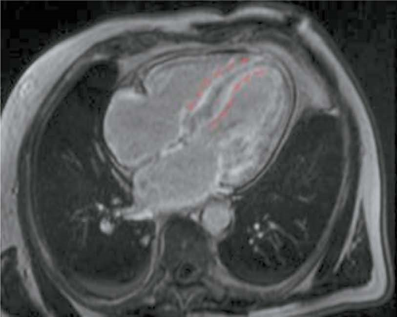
Potential use of cardiac magnetic resonance imaging in differential diagnosis of cardiomyopathies due to light-chain amyloidosis and transthyretin amyloidosis
- Authors: Magomedova Z.M.1,2, Nikiforova T.V.3, Shchekochikhin D.Y.1,2, Pershina E.S.1,2, Kovalev K.V.1, Abdulmazhidova K.S.2, Rassechkina D.S.2, Grachev A.E.4, Rekhtina I.G.4, Sarkisyan S.D.2, Volovchenko A.N.2, Sinitsyn V.E.5, Andreev D.A.2
-
Affiliations:
- Pirogov Municipal Clinical Hospital № 1
- Sechenov First Moscow State Medical University
- S.S. Yudin City Clinical Hospital
- National Medical Research Center of Hematology
- Lomonosov Moscow State University
- Issue: Vol 5, No 4 (2024)
- Pages: 668-681
- Section: Original Study Articles
- URL: https://bakhtiniada.ru/DD/article/view/309828
- DOI: https://doi.org/10.17816/DD635007
- ID: 309828
Cite item
Abstract
BACKGROUND: Cardiac amyloidosis is a serious progressive disease with a high mortality rate. The differential diagnosis of cardiomyopathies due to amyloid light-chain (AL) amyloidosis and transthyretin (ATTR) amyloidosis is important for selecting the optimal treatment strategy.
AIM: The aim of this study was to evaluate the capabilities of cardiac magnetic resonance imaging in the differential diagnosis of cardiomyopathies due to AL and ATTR amyloidosis.
MATERIALS AND METHODS: A retrospective analysis of the medical records of 25 patients with a confirmed diagnosis of amyloid cardiomyopathy was performed. Patients were divided into two groups according to the type of amyloidosis, with group 1 including patients with cardiomyopathy due to AL amyloidosis and group 2 including patients with cardiomyopathy due to ATTR amyloidosis. All patients underwent contrast-enhanced cardiac magnetic resonance imaging. Volumetric and linear cardiac parameters, ventricular function, and late gadolinium enhancement patterns were assessed. Standard statistical methods were used, and differences were considered significant at p <0.05.
RESULTS: Group 2 showed a more significant thickening of the myocardial walls compared to group 1 (interventricular septum: 18 [17; 18] vs. 14.5 mm [12.8; 16.0], p <0.01, posterior wall of the left ventricle: 14 [13; 17] vs. 10.5 mm [10; 12.3], p <0.01). The indexed mass of the left ventricle myocardium was 110 [92; 125] in group 2 and 85 mm [69.3; 91.8] in group 1 (p <0.01). In group 2, late gadolinium enhancement with a transmural left ventricle pattern was more frequently observed in the basal and mid-lower-lateral segments, whereas in group 1, a subendocardial pattern of late gadolinium enhancement was more frequent in the mid-anterior and lower-lateral segments (p <0.05). In addition, frequency of simultaneous contrast enhancement in the subendocardial layers of the interventricular septum on the left ventricle and right ventricle sides was higher in group 2 (100% of cases vs. 50%, p <0.01). Late gadolinium enhancement of the right ventricle was also more common in group 2 (100 vs. 58%, p <0.05), especially in the interventricular septum and inferior wall area (p <0.05). Semi-quantitative assessment of LGE using the Query Amyloid Late Enhancement (QALE) showed greater contrast enhancement in group 2: 13 [12; 14] vs. 10.5 [1.75; 12], p <0.01), and a score greater than 13 differentiated between cardiomyopathy due to AL amyloidosis and ATTR amyloidosis with a sensitivity of 69% and a specificity of 83%.
CONCLUSIONS: Cardiac MRI identifies typical features of cardiomyopathies due to AL amyloidosis and ATTR amyloidosis for their differential diagnosis. Further research is needed to confirm diagnostic accuracy of the patterns identified.
Full Text
##article.viewOnOriginalSite##About the authors
Zainab M. Magomedova
Pirogov Municipal Clinical Hospital № 1; Sechenov First Moscow State Medical University
Author for correspondence.
Email: magomedova.zainab.97@mail.ru
ORCID iD: 0000-0001-6753-1525
SPIN-code: 5271-4915
MD, radiologist of the Department of Magnetic Resonance and Computed Tomography of the Clinical Hospital No. 1 named after N.I. Pirogov; postgraduate student of the Cardiology Department, Functional and Ultrasound Diagnostics at I.M. Sechenov Moscow State Medical University (Sechenov University).
Russian Federation, Moscow; MoscowTatyana V. Nikiforova
S.S. Yudin City Clinical Hospital
Email: attrcmp@gmail.com
ORCID iD: 0000-0003-3072-8951
SPIN-code: 4997-0330
MD, cardiologist
Russian Federation, MoscowDmitry Y. Shchekochikhin
Pirogov Municipal Clinical Hospital № 1; Sechenov First Moscow State Medical University
Email: agishm@list.ru
ORCID iD: 0000-0002-8209-2791
SPIN-code: 3753-6915
MD, Cand. Sci. (Medicine), Assistant Professor
Russian Federation, Moscow; MoscowEkaterina S. Pershina
Pirogov Municipal Clinical Hospital № 1; Sechenov First Moscow State Medical University
Email: pershina86@mail.ru
ORCID iD: 0000-0002-3952-6865
SPIN-code: 7311-9276
MD, Cand. Sci. (Medicine), Deputy Chief Physician for or Strategic Development and Head, Associate Professor of the Department of Cardiology, Functional and Ultrasound Diagnostics), Senior Researcher at the Institute of Personalized Cardiology
Russian Federation, Moscow; MoscowKonstantin V. Kovalev
Pirogov Municipal Clinical Hospital № 1
Email: radix606@yandex.ru
ORCID iD: 0009-0004-4841-041X
MD, radiologist of the Department of Magnetic Resonance and Computed Tomography
Russian Federation, MoscowKhadizhat S. Abdulmazhidova
Sechenov First Moscow State Medical University
Email: abdulmazhidova.kh@mail.ru
ORCID iD: 0009-0008-5064-7802
student
Russian Federation, MoscowDaria S. Rassechkina
Sechenov First Moscow State Medical University
Email: rassechkina@yandex.ru
ORCID iD: 0009-0007-8825-8485
MD, resident of the Department of Cardiology, Functional and Ultrasound Diagnostics
Russian Federation, MoscowAlexander E. Grachev
National Medical Research Center of Hematology
Email: gra4al@yandex.ru
ORCID iD: 0000-0001-7221-9392
SPIN-code: 4281-3923
MD, Cand. Sci. (Medicine), Hematologist
Russian Federation, MoscowIrina G. Rekhtina
National Medical Research Center of Hematology
Email: rekhtina.i@blood.ru
ORCID iD: 0000-0002-7944-6202
SPIN-code: 4920-7144
MD, Dr. Sci. (Medicine), Head of the Department of Hematology and Chemotherapy of Plasma Cell Dyscrasias, Hematologist
Russian Federation, MoscowSusanna D. Sarkisyan
Sechenov First Moscow State Medical University
Email: sysanna.sarkisyan.2001@mail.ru
ORCID iD: 0000-0002-6454-1370
student
Russian Federation, MoscowAlexey N. Volovchenko
Sechenov First Moscow State Medical University
Email: dr.volovchenko@mail.ru
ORCID iD: 0000-0002-0923-735X
SPIN-code: 4120-8740
MD, Cand. Sci. (Medicine), Head of the Cardiology Department at the Cardiology Clinic, Assistant Professor at the Department of Cardiology, Functional and Ultrasound Diagnostics
Russian Federation, MoscowValentin E. Sinitsyn
Lomonosov Moscow State University
Email: vsini@mail.ru
ORCID iD: 0000-0002-5649-2193
SPIN-code: 8449-6590
MD, Dr. Sci. (Medicine), Professor, Head of the Department of Radiology and Therapy, Head of the Department of Radiology at the Faculty of Fundamental Medicine and the Interdisciplinary Scientific and Educational School
Russian Federation, MoscowDenis A. Andreev
Sechenov First Moscow State Medical University
Email: dennan@mail.ru
ORCID iD: 0000-0002-0276-7374
SPIN-code: 8790-8834
MD, Dr. Sci. (Medicine), Head of the Department of Cardiology, Functional and Ultrasound Diagnostics
Russian Federation, MoscowReferences
- Wechalekar AD, Gillmore JD, Hawkins PN. Systemic amyloidosis. Lancet. 2016;387(10038):2641–2654. doi: 10.1016/S0140-6736(15)01274-X
- Rapezzi C, Lorenzini M, Longhi S, et al. Cardiac amyloidosis: the great pretender. Heart Fail Rev. 2015;20(2):117–124. doi: 10.1007/s10741-015-9480-0
- Myasnikov RP, Andreyenko EYu, Kushunina DV, et al. Cardiac amyloidosis: modern aspects of diagnosis and treatment (clinical observation). Clinical and experimental surgery. 2014;(4):72–82. EDN: TRLZYN
- Maurer MS, Elliott P, Comenzo R, et al. Addressing common questions encountered in the diagnosis and management of cardiac amyloidosis. Circulation. 2017;135(14):1357–1377. doi: 10.1161/CIRCULATIONAHA.116.024438
- Ruberg FL, Grogan M, Hanna M, et al. Transthyretin amyloid cardiomyopathy: JACC state of the art review. J Am Coll Cardiol. 2019;73(22):2872–2891. doi: 10.1016/j.jacc.2019.04.003
- Kittleson MM, Maurer MS, Ambardekar AV, et al.; American Heart Association Heart Failure and Transplantation Committee of the Council on Clinical Cardiology. Cardiac amyloidosis: evolving diagnosis and management: a scientific statement from the American Heart Association. Circulation. 2020;142(1):e7–e22. doi: 10.1161/CIR.0000000000000792
- Maurer MS, Bokhari S, Damy T, et al. Expert consensus recommendations for the suspicion and diagnosis of transthyretin cardiac amyloidosis. Circ Heart Fail. 2019;12(9):e006075. doi: 10.1161/CIRCHEARTFAILURE.119.006075
- Garcia Pavia P, Rapezzi C, Adler Y, et al. Diagnosis and treatment of cardiac amyloidosis: a position statement of the ESC Working Group on Myocardial and Pericardial Diseases. Eur Heart J. 2021;42(16):1554–1568. doi: 10.1093/eurheartj/ehab072
- Lysenko LV, Rameev VV, Moiseev SV, et al. Clinical guidelines for diagnosis and treatment of systemic amyloidosis. Clinical pharmacology and therapy. 2020;29(1):13–24. EDN UCEZAB doi: 10.32756/ 0869-5490-2020-1-13-24
- Syed IS, Glockner JF, Feng D, et al. Role of cardiac magnetic resonance imaging in the detection of cardiac amyloidosis. JACC Cardiovasc Imaging. 2010;3(2):155–164. doi: 10.1016/j.jcmg.2009.09.023
- Butorova EA, Stukalova OV. Role of cardiac MRI in the diagnosis of cardiac amyloidosis. Clinical cases. Clinical review for general practice. 2021;(2):16–20. EDN MUWTXO doi: 10.47407/kr2021.2.2.00037
- Dorbala S, Ando Y, Bokhari S, et al. ASNC/AHA/ASE/EANM/HFSA/ISA/SCMR/SNMMI expert consensus recommendations for multimodality imaging in cardiac amyloidosis: Part 1 of 2-evidence base and standardized methods of imaging. J Nucl Cardiol. 2019;26(6):2065–2123. doi: 10.1007/s12350-019-01760-6
- Dungu JN, Valencia O, Pinney JH, et al. CMR-based differentiation of AL and ATTR cardiac amyloidosis. JACC Cardiovasc Imaging. 2014;7(2):133–142. doi: 10.1016/j.jcmg.2013.08.015
- Itzhaki Ben Zadok O, Vaturi M, Vaxman I, et al. Differences in the characteristics and contemporary cardiac outcomes of patients with light chain versus transthyretin cardiac amyloidosis. PLoS One. 2021;16(8):e0255487. doi: 10.1371/journal.pone.0255487
- Quarta CC, Solomon SD, Uraizee I, et al. Left ventricular structure and function in transthyretin related versus light chain cardiac amyloidosis. Circulation. 2014;129(18):1840–1849. doi: 10.1161/CIRCULATIONAHA.113.006242
- Stern LK, Patel J. Cardiac Amyloidosis Treatment. Methodist Debakey Cardiovasc J. 2022;18(2):59–72. doi: 10.14797/mdcvj.1050
- Kristen AV., aus dem Siepen F, Scherer K, et al. Comparison of different types of cardiac amyloidosis by cardiac magnetic resonance imaging. Amyloid. 2015;22(2):132–141. doi: 10.3109/13506129.2015.1020153
- Gillmore JD, Maurer MS, Falk RH, et al. Nonbiopsy diagnosis of cardiac transthyretin amyloidosis. Circulation. 2016;133(24):2404–2412. doi: 10.1161/CIRCULATIONAHA.116.021612
- Martinez Naharro A, Treibel TA, Abdel Gadir A, et al. Magnetic resonance in transthyretin cardiac amyloidosis. J Am Coll Cardiol. 2017;70(4):466–477. doi: 10.1016/j.jacc.2017.05.053
- Binder C, Duca F, Binder T, et al. Prognostic implications of pericardial and pleural effusion in patients with cardiac amyloidosis. Clinical Research in Cardiology. 2021;110(4):532–543. doi: 10.1007/s00392-020-01698-7
- Reddy A, Singh V, Karthikeyan B, et al. Biventricular strain imaging with cardiac MRI in genotyped and histology validated amyloid cardiomyopathy. Cardiogenetics. 2021;11(3):98–110. doi: 10.3390/cardiogenetics11030011
Supplementary files


