

")


Mitral valve calcinosis as an important finding during heart examination
- Authors: Filatova D.A.1, Mershina E.A.1, Plotnikova M.L.1, Lisitskaya M.V.1, Sinitsyn V.E.1
-
Affiliations:
- Lomonosov Moscow State University
- Issue: Vol 5, No 2 (2024)
- Pages: 219-230
- Section: Original Study Articles
- URL: https://bakhtiniada.ru/DD/article/view/264834
- DOI: https://doi.org/10.17816/DD624754
- ID: 264834
Cite item

Abstract
BACKGROUND: Mitral valve calcinosis is a chronic degenerative process in the fibrous structures of the mitral valve. Advanced stages increase the risk of endocarditis and cardiac rhythm disturbances and contribute to cardiovascular mortality. The cause of mitral valve calcinosis is still controversial; however, the contribution of atherosclerosis to its development is currently undisputed. The prevalence of mitral valve calcinosis varies in different age groups and on average is higher in people with cardiovascular disease.
AIM: To assess the prevalence of mitral valve calcinosis in patients undergoing computed tomography angiography and identify the relationship between aortic and mitral valve calcinosis and coronary calcium index and signs of remodeling.
MATERIALS AND METHODS: A retrospective study of 336 patients who underwent computed tomography coronary angiography with intravenous contrast enhancement at the Lomonosov Moscow State University Clinic between November 13, 2020, and May 14, 2022, was conducted.
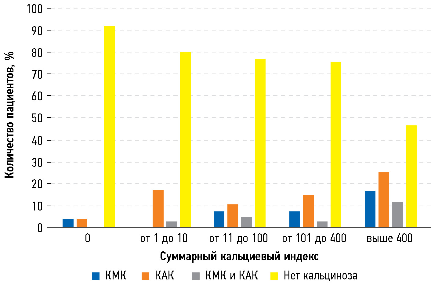
RESULTS: The prevalence of aortic (16.4%) and mitral (11%) valve calcinosis was high in people undergoing cardiovascular examination, and a relationship was noted between valve calcinosis and coronary calcium index.
CONCLUSION: The detection of mitral valve calcinosis in patients during routine examination is important in predicting further treatment and outcomes because valve calcinosis is an indirect indicator of coronary heart disease risk. Although valve calcinosis is usually an incidental examination finding, it may indicate a high cardiovascular risk and should prompt further evaluation, if clinically necessary.
Full Text
##article.viewOnOriginalSite##About the authors
Daria A. Filatova
Lomonosov Moscow State University
Author for correspondence.
Email: dariafilatova.msu@mail.ru
ORCID iD: 0000-0002-0894-1994
SPIN-code: 2665-5973
MD
Russian Federation, MoscowElena A. Mershina
Lomonosov Moscow State University
Email: elena_mershina@mail.ru
ORCID iD: 0000-0002-1266-4926
SPIN-code: 6897-9641
MD, Cand. Sci. (Medicine)
Russian Federation, MoscowMaria L. Plotnikova
Lomonosov Moscow State University
Email: maria_plotnikova@inbox.ru
ORCID iD: 0000-0001-7533-9867
SPIN-code: 1857-0770
MD
Russian Federation, MoscowMariya V. Lisitskaya
Lomonosov Moscow State University
Email: lissenok@inbox.ru
ORCID iD: 0000-0002-8402-7643
SPIN-code: 2301-8480
MD, Cand. Sci. (Medicine)
Russian Federation, MoscowValentin E. Sinitsyn
Lomonosov Moscow State University
Email: vsini@mail.ru
ORCID iD: 0000-0002-5649-2193
SPIN-code: 8449-6590
MD, Dr. Sci. (Medicine), Professor
Russian Federation, MoscowReferences
- Maher ER, Young G, Smyth-Walsh B, Pugh S, Curtis JR. Aortic and mitral valve calcification in patients with end-stage renal disease. Lancet. 1987;330(8564):875–877. doi: 10.1016/s0140-6736(87)91370-5
- Fox E, Harkins D, Taylor H, et al. Epidemiology of mitral annular calcification and its predictive value for coronary events in African Americans: the Jackson Cohort of the Atherosclerotic Risk in Communities Study. Am. Heart J. 2004;148(6):979–984. doi: 10.1016/j.ahj.2004.05.048
- Atar S, Jeon DS, Luo H, Siegel RJ. Mitral annular calcification: a marker of severe coronary artery disease in patients under 65 years old. Heart. 2003;89(2):161–164. doi: 10.1136/heart.89.2.161
- Savage DD, Garrison RJ, Castelli WP, et al. Prevalence of submitral (anular) calcium and its correlates in a general population-based sample (the Framingham Study). Am. J. Cardiol. 1983;51(8):1375–1378. doi: 10.1016/0002-9149(83)90315-6
- Barasch E, Gottdiener JS, Larsen EKM, et al. Clinical significance of calcification of the fibrous skeleton of the heart and aortosclerosis in community dwelling elderly. The Cardiovascular Health Study (CHS). Am. Heart J. 2006;151(1):39–47. doi: 10.1016/j.ahj.2005.03.052
- Nestico PF, Depace NL, Morganroth J, Kotler MN, Ross J. Mitral annular calcification: clinical, pathophysiology, and echocardiographic review. Am. Heart J. 1984;107(5 Pt 1):989–996. doi: 10.1016/0002-8703(84)90840-8
- Stary HC, Blankenhorn DH, Chandler AB, et al. A definition of the intima of human arteries and of its atherosclerosis-prone regions. A report from the Committee on Vascular Lesions of the Council on Arteriosclerosis, American Heart Association. Arterioscler. Thromb. J. Vasc. Biol. 1992;12(1):120–134. doi: 10.1161/01.atv.12.1.120
- Allison MA, Cheung P, Criqui MH, Langer RD, Wright CM. Mitral and Aortic Annular Calcification Are Highly Associated With Systemic Calcified Atherosclerosis. Circulation. 2006;113(6):861–866. doi: 10.1161/CIRCULATIONAHA.105.552844
- Otto CM, Kuusisto J, Reichenbach DD, Gown AM, O’Brien KD. Characterization of the early lesion of “degenerative” valvular aortic stenosis. Histological and immunohistochemical studies. Circulation. 1994;90(2):844–853. doi: 10.1161/01.cir.90.2.844
- Mohler ER. Mechanisms of aortic valve calcification. Am. J. Cardiol. 2004;94(11):1396–1402. doi: 10.1016/j.amjcard.2004.08.013
- Shahi CN, Ghaisas NK, Goggins M, et al. Elevated levels of circulating soluble adhesion molecules in patients with nonrheumatic aortic stenosis. Am. J. Cardiol. 1997;79(7):980–982. doi: 10.1016/s0002-9149(97)00027-1
- Olsson M, Thyberg J, Nilsson J. Presence of oxidized low density lipoprotein in nonrheumatic stenotic aortic valves. Arterioscler. Thromb. Vasc. Biol. 1999;19(5):1218–1222. doi: 10.1161/01.atv.19.5.1218
- Edep ME, Shirani J, Wolf P, Brown DL. Matrix metalloproteinase expression in nonrheumatic aortic stenosis. Cardiovasc. Pathol. 2000;9(5):281–286. doi: 10.1016/s1054-8807(00)00043-0
- O’Brien KD, Shavelle DM, Caulfield MT, et al. Association of Angiotensin-Converting Enzyme With Low-Density Lipoprotein in Aortic Valvular Lesions and in Human Plasma. Circulation. 2002;106(17):2224–2230. doi: 10.1161/01.CIR.0000035655.45453.D2
- Pohle K, Otte M, Mäffert R, et al. Association of cardiovascular risk factors to aortic valve calcification as quantified by electron beam computed tomography. Mayo Clin. Proc. 2004;79(10):1242–1246. doi: 10.4065/79.10.1242
- Wong ND, Sciammarella M, Arad Y, et al. Relation of thoracic aortic and aortic valve calcium to coronary artery calcium and risk assessment. Am. J. Cardiol. 2003;92(8):951–955. doi: 10.1016/s0002-9149(03)00976-7
- Fox CS, Vasan RS, Parise H, et al. Mitral Annular Calcification Predicts Cardiovascular Morbidity and Mortality. Circulation. 2003;107(11):1492–1496. doi: 10.1161/01.CIR.0000058168.26163.BC
- Tenenbaum A, Fisman EZ, Pines A, et al. Gender paradox in cardiac calcium deposits in middle-aged and elderly patients: mitral annular and coronary calcifications interrelationship. Maturitas. 2000;36(1):35–42. doi: 10.1016/s0378-5122(00)00120-1
- Sugihara N, Matsuzaki M. The influence of severe bone loss on mitral annular calcification in postmenopausal osteoporosis of elderly Japanese women. Jpn. Circ. J. 1993;57(1):14–26. doi: 10.1253/jcj.57.14
- Elmariah S, Delaney JAC, O’Brien KD, et al. Bisphosphonate Use and Prevalence of Valvular and Vascular Calcification in Women MESA (The Multi-Ethnic Study of Atherosclerosis). J. Am. Coll. Cardiol. 2010;56(21):1752–1759. doi: 10.1016/j.jacc.2010.05.050
- Elmariah S, Delaney JAC, Bluemke DA, et al. Associations of LV hypertrophy with prevalent and incident valve calcification: Multi-Ethnic Study of Atherosclerosis. JACC Cardiovasc. Imaging. 2012;5(8):781–788. doi: 10.1016/j.jcmg.2011.12.025
- Adler Y, Koren A, Fink N, et al. Association between mitral annulus calcification and carotid atherosclerotic disease. Stroke. 1998;29(9):1833–1837. doi: 10.1161/01.str.29.9.1833
- Umana E, Ahmed W, Alpert MA. Valvular and perivalvular abnormalities in end-stage renal disease. Am. J. Med. Sci. 2003;325(4):237–242. doi: 10.1097/00000441-200304000-00010
- Alfrey AC. The role of abnormal phosphorus metabolism in the progression of chronic kidney disease and metastatic calcification. Kidney Int. Suppl. 2004;(90):S13–S17. doi: 10.1111/j.1523-1755.2004.09003.x
- Jesri A, Braitman LE, Pressman GS. Severe mitral annular calcification predicts chronic kidney disease. Int. J. Cardiol. 2008;128(2):193–196. doi: 10.1016/j.ijcard.2007.05.015
- Ribeiro S, Ramos A, Brandão A, et al. Cardiac valve calcification in haemodialysis patients: role of calcium-phosphate metabolism. Nephrol. Dial. Transplant. 1998;13(8):2037–2040. doi: 10.1093/ndt/13.8.2037
- Correia J, Rodrigues D, da Silva AM, Sá e Melo A, Providência LA. Massive calcification of the mitral valve annulus in an adolescent with Marfan syndrome. A case report. Rev. Port. Cardiol. 2006;25(10):921–926.
- Völzke H, Haring R, Lorbeer R, et al. Heart valve sclerosis predicts all-cause and cardiovascular mortality. Atherosclerosis. 2010;209(2):606–610. doi: 10.1016/j.atherosclerosis.2009.10.030
- Tenenbaum A, Shemesh J, Fisman EZ, Motro M. Advanced mitral annular calcification is associated with severe coronary calcification on fast dual spiral computed tomography. Invest. Radiol. 2000;35(3):193–198. doi: 10.1097/00004424-200003000-00006
- Kizer JR, Wiebers DO, Whisnant JP, et al. Mitral annular calcification, aortic valve sclerosis, and incident stroke in adults free of clinical cardiovascular disease: the Strong Heart Study. Stroke. 2005;36(12):2533–2537. doi: 10.1161/01.STR.0000190005.09442.ad
- Rodriguez CJ, Bartz TM, Longstreth WT, et al. Association of annular calcification and aortic valve sclerosis with brain findings on magnetic resonance imaging in community dwelling older adults: the cardiovascular health study. J. Am. Coll. Cardiol. 2011;57(21):2172–2180. doi: 10.1016/j.jacc.2011.01.034
- O’Neal WT, Efird JT, Nazarian S, et al. Mitral annular calcification and incident atrial fibrillation in the Multi-Ethnic Study of Atherosclerosis. EP Europace. 2015;17(3):358–363. doi: 10.1093/europace/euu265
- Willens HJ, Ferreira AC, Gallagher AJ, Morytko JA. Mobile components associated with rapidly developing mitral annulus calcification in patients with chronic renal failure: review of mobile elements associated with mitral annulus calcification. Echocardiogr. 2003;20(4):363–367. doi: 10.1046/j.1540-8175.2003.03042.x
- Movahed MR, Saito Y, Ahmadi-Kashani M, Ebrahimi R. Mitral Annulus Calcification is associated with valvular and cardiac structural abnormalities. Cardiovasc. Ultrasound. 2007;5(1):14. doi: 10.1186/1476-7120-5-14
- Vistarini N, d’Alessandro C, Aubert S, et al. Surgery for infective endocarditis on mitral annulus calcification. J. Heart Valve Dis. 2007;16(6):611–616.
- Fulkerson PK, Beaver BM, Auseon JC, Graber HL. Calcification of the mitral annulus: Etiology, clinical associations, complications and therapy. Am. J. Med. 1979;66(6):967–977. doi: 10.1016/0002-9343(79)90452-2
- Takamoto T, Popp RL. Conduction disturbances related to the site and severity of mitral anular calcification: A 2-dimensional echocardiographic and electrocardiographs correlative study. Am. J. Cardiol. 1983;51(10):1644–1649. doi: 10.1016/0002-9149(83)90202-3
- Pekdemir H, Cansel M, Yağmur J, et al. Assessment of atrial conduction time by tissue Doppler echocardiography and P-wave dispersion in patients with mitral annulus calcification. J. Electrocardiol. 2010;43(4):339–343. doi: 10.1016/j.jelectrocard.2010.02.013
- Sveric KM, Platzek I, Golgor E, et al. Purposeful use of multimodality imaging in the diagnosis of caseous mitral annular calcification: a case series report. BMC Med. Imaging. 2022;22:7. doi: 10.1186/s12880-021-00725-x
- Tyebally S, Chen D, Bhattacharyya S, et al. Cardiac Tumors: JACC CardioOncology State-of-the-Art Review. JACC CardioOncology. 2020;2(2):293–311. doi: 10.1016/j.jaccao.2020.05.009
- Mayr A, Müller S, Feuchtner G. The Spectrum of Caseous Mitral Annulus Calcifications. JACC Case Rep. 2020;3(1):104–108. doi: 10.1016/j.jaccas.2020.09.039
Supplementary files


