

")


Conventional structural magnetic resonance imaging in differentiating chronic disorders of consciousness
- Authors: Sergeeva A.N.1, Morozova S.N.1, Sergeev D.V.1, Kremneva E.I.1, Zimin A.A.1, Legostaeva L.A.1, Iazeva E.G.2, Krotenkova M.V.1, Ryabinkina Y.V.1, Suponeva N.A.1, Piradov M.A.1
-
Affiliations:
- Research Center of Neurology
- LLC “Three sisters” Rehabilitation center
- Issue: Vol 5, No 2 (2024)
- Pages: 190-202
- Section: Original Study Articles
- URL: https://bakhtiniada.ru/DD/article/view/264832
- DOI: https://doi.org/10.17816/DD569418
- ID: 264832
Cite item

Abstract
BACKGROUND: Differential diagnosis of chronic disorders of consciousness remains one of the most difficult problems even for experienced clinicians.
AIM: To evaluate the inter-expert consistency and capacity of the researcher-developed structural scale based on magnetic resonance imaging to differentiate chronic disorders of consciousness, named, DOC-MRIDS, on a larger sample of patients.
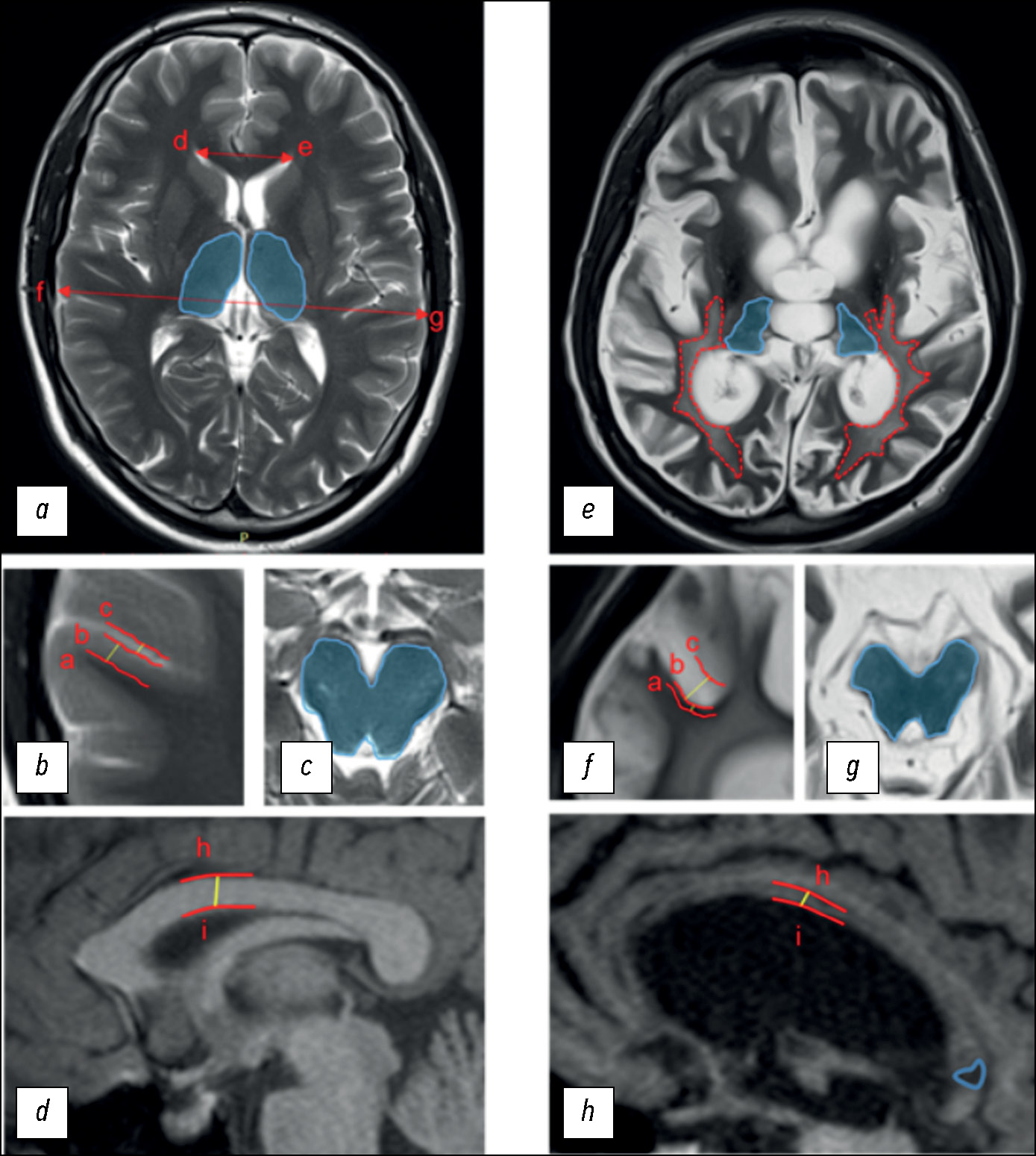
MATERIALS AND METHODS: Sixty patients with a clinically stable status diagnosed with consciousness disorders (vegetative state, n=32; minimally conscious state, n=28) were enrolled. The revised coma recovery scale (CRS-R) was included in the clinical assessment. All patients underwent structural magnetic resonance imaging with 3.0-T Siemens scanners including T2 and T1 sequences. Structural changes were assessed using the DOC-MRIDS scale and included the following features: diffuse cortical atrophy, ventricular enlargement, gyri dilatation, leukoaraiosis, brainstem and/or thalamic degeneration, corpus callosum degeneration, and focal corpus callosum lesions. A total score was calculated. Magnetic resonance imaging data were analyzed by three neuroradiologists, and inter-observer agreement (Krippendorf’s alpha) was assessed.
RESULTS: A high inter-examiner agreement of the DOC-MRIDS scale score was found, with α=0.806 (95% confidence interval 0.757–0.849). The vegetative state group had a higher DOC-MRIDS score than the minimally conscious state group (p <0.005). A negative correlation was obtained between CRS-R and DOC-MRIDS scale scores (ρ=–0.457, p <0.0001), individual clinical scale domains, and magnetic resonance imaging features.
CONCLUSION: When assessing structural changes in patients with chronic consciousness disorders, the use of the DOC-MRIDS scale helps differentiate the type of such disorders with sufficient specificity, sensitivity, and inter-rater agreement. This scale can be used in clinical practice as an additional differential diagnostic tool.
Full Text
##article.viewOnOriginalSite##About the authors
Anastasia N. Sergeeva
Research Center of Neurology
Author for correspondence.
Email: sergeeva@neurology.ru
ORCID iD: 0000-0002-2481-4565
SPIN-code: 6761-8250
MD, Cand. Sci. (Medicine)
Russian Federation, MoscowSofya N. Morozova
Research Center of Neurology
Email: kulikovasn@gmail.com
ORCID iD: 0000-0002-9093-344X
SPIN-code: 2434-7827
MD, Cand. Sci. (Medicine)
Russian Federation, MoscowDmitrii V. Sergeev
Research Center of Neurology
Email: dmsergeev@yandex.ru
ORCID iD: 0000-0002-9130-1292
SPIN-code: 8282-3920
MD, Cand. Sci. (Medicine)
Russian Federation, MoscowElena I. Kremneva
Research Center of Neurology
Email: moomin10j@mail.ru
ORCID iD: 0000-0001-9396-6063
SPIN-code: 8799-8092
MD, Cand. Sci. (Medicine)
Russian Federation, MoscowAlexey A. Zimin
Research Center of Neurology
Email: leha-zimin@inbox.ru
ORCID iD: 0000-0002-9226-2870
SPIN-code: 9525-1805
Russian Federation, Moscow
Lyudmila A. Legostaeva
Research Center of Neurology
Email: milalegostaeva@gmail.com
MD, Cand. Sci. (Medicine)
Russian Federation, MoscowElizaveta G. Iazeva
LLC “Three sisters” Rehabilitation center
Email: lizaveta.mochalova@gmail.com
ORCID iD: 0000-0003-0382-7719
SPIN-code: 4895-3900
MD, Cand. Sci. (Medicine)
Russian Federation, MoscowMarina V. Krotenkova
Research Center of Neurology
Email: krotenkova_mrt@mail.ru
ORCID iD: 0000-0003-3820-4554
SPIN-code: 9663-8828
MD, Dr. Sci. (Medicine)
Russian Federation, МоскваYulia V. Ryabinkina
Research Center of Neurology
Email: ryabinkina11@mail.ru
ORCID iD: 0000-0001-8576-9983
SPIN-code: 5044-2701
MD, Dr. Sci. (Medicine)
Russian Federation, MoscowNatalya A. Suponeva
Research Center of Neurology
Email: nasu2709@mail.ru
ORCID iD: 0000-0003-3956-6362
SPIN-code: 3223-6006
MD, Dr. Sci. (Medicine), corresponding member of the Russian Academy of Sciences, Professor
Russian Federation, MoscowMichael A. Piradov
Research Center of Neurology
Email: mpi711@gmail.com
ORCID iD: 0000-0002-6338-0392
SPIN-code: 2860-1689
MD, Dr. Sci. (Medicine), academician member of the Russian Academy of Sciences, Professor
Russian Federation, MoscowReferences
- Koch C, Massimini M, Boly M, Tononi G. Neural correlates of consciousness: progress and problems. Nat Rev Neurosci. 2016;17(5):307–321. doi: 10.1038/nrn.2016.22
- Monti MM, Laureys S, Owen AM. The vegetative state. BMJ. 2010;341:376–385. doi: 10.1136/bmj.c3765
- Giacino JT, Ashwal S, Childs N, et al. The minimally conscious state: Definition and diagnostic criteria. Neurology. 2002;58(3):349–353. doi: 10.1212/wnl.58.3.349
- Belkin AA, Aleksandrova EV, Akhutina TV, et al. Chronic Disorders of Consciousness: guidelines of the All-Russian public organization “Federation of Anesthesiologists and Reanimatologists”. Annals of critical care. 2023;(3):7–42. doi: 10.21320/1818-474X-2023-3-7-42
- Giacino JT. The vegetative and minimally conscious states: Consensus-based criteria for establishing diagnosis and prognosis. Neurorehabilitation. 2004;19(4):293–298. doi: 10.3233/NRE-2004-19405
- Seel RT, Sherer M, et al. Assessment scales for disorders of consciousness: evidence-based recommendations for clinical practice and research. Arch Phys Med Rehabil. 2010;91(12):1795–1813. doi: 10.1016/j.apmr.2010.07.218
- Schnakers C, Vanhaudenhuyse A, Giacino J, et al. Diagnostic accuracy of the vegetative and minimally conscious state: Clinical consensus versus standardized neurobehavioral assessment. BMC Neurol. 2009;(9):35–40. doi: 10.1186/1471-2377-9-35
- Stender J, Gosseries O, Bruno M, et al. Diagnostic precision of PET imaging and functional MRI in disorders of consciousness: A clinical validation study. Lancet. 2014;384(9942):514–522. doi: 10.1016/S0140-6736(14)60042-8
- Monti M, Vanhaudenhuyse A, Coleman M, et al. Willful modulation of brain activity in disorders of consciousness. N. Engl. J. Med. 2010;362(7):579–589. doi: 10.1056/NEJMoa0905370
- Crone J, Bio B, Vespa P, et. al. Restoration of thalamo-cortical connectivity after brain injury: Recovery of consciousness, complex behavior, or passage of time. J. Neurosci. Res. 2018;96(4):671–687. doi: 10.1002/jnr.24115
- Demertzi A, Antonopoulos G, Heine L, et al. Intrinsic functional connectivity differentiates minimally conscious from unresponsive patients. Brain. 2015;138(9):2619–2631. doi: 10.1093/brain/awv169
- Lutkenhoff E, Chiang J, Tshibanda L, et al. Thalamic and extrathalamic mechanisms of consciousness after severe brain injury. Ann. Neurol. 2015;78(1):68–76. doi: 10.1002/ana.24423
- Guldenmund P, Soddu A, Baquero K, et al. Structural brain injury in patients with disorders of consciousness: A voxel-based morphometry study. Brain Inj. 2016;30(3):343–352. doi: 10.3109/02699052.2015.1118765
- Annen J, Frasso G, Crone J, et al. Regional brain volumetry and brain function in severely brain-injured patients. Ann. Neurol. 2018;83(4):842–853. doi: 10.1002/ana.25214
- Morozova SN, Kremneva EI, Sergeev DV, et al. Conventional Structural Magnetic Resonance Imaging in Differentiating Chronic Disorders of Consciousness. Brain Sci. 2018;8(8):144–155. doi: 10.3390/brainsci8080144
- Legostaeva LA, Mochalova EG, Suponeva NA, et al. Difficulties in evaluation of chronic disorders of consciousness: approaches to clinical assessment and instrumental studies. Russian Journal of Anesthesiology and Reanimatology. 2017;62(6):449–456. EDN: YPLNJY doi: 10.18821/0201-7563-2017-62-6-449-456
- Solovyeva PI, Sinkin МV, Talypov АE, et al. Clinical assessment of patients with chronic disorders of consciousness by different medical specialists. Annals of Clinical and Experimental Neurology. 2022;16(2):44–49. doi: 10.54101/ACEN.2022.2.5
- Medina JP, Nigri A, Stanziano M, et al. Resting-State fMRI in Chronic Patients with Disorders of Consciousness: The Role of Lower-Order Networks for Clinical Assessment. Brain Sci. 2022;12(3):355–374. doi: 10.3390/brainsci12030355
- Rohaut B, Doyle KW, Reynolds AS, et al. Deep structural brain lesions associated with consciousness impairment early after hemorrhagic stroke. Sci Rep. 2019;9(1):4174. doi: 10.1038/s41598-019-41042-2
- Alnagger N, Cardone P, Martial C, et al. The current and future contribution of neuroimaging to the understanding of disorders of consciousness. Presse Med. 2023;52(2):104163. doi: 10.1016/j.lpm.2022.104163
- Bakulin IS, Kremneva EI, Kuznetsov AV, et al. Chronic disorders of consciousness. Piradov MA, editor. Moscow: Hot line — Telecom; 2020.
Supplementary files


