


")


Intestinal Dysfunction in Acute Peritonitis
- Authors: Al-Kubaisi S.S.1, Al-Mihyawi F.M.1, Abed M.A.1, Al-Mashhadani A.A.1, Younus A.R.1, Al-Jizani H.A.1, Dhari L.H.1
-
Affiliations:
- National Research Mordovia State University
- Issue: Vol 1, No 3 (2025)
- Pages: 224-231
- Section: Pathological physiology
- Submitted: 08.05.2025
- Accepted: 12.09.2025
- Published: 22.09.2025
- URL: https://bakhtiniada.ru/3034-6231/article/view/290779
- DOI: https://doi.org/10.15507/3034-6231.001.202503.224-231
- EDN: https://elibrary.ru/pojszp
- ID: 290779

Cite item
Full Text

Abstract
Introduction. Acute peritonitis continues to be a significant medical issue because of its high mortality, especially in the terminal phase. The aim of this research is to define the role of lipid peroxidation processes in the disruption of small bowel function among patients with acute peritonitis.
Materials and methods. A clinical study included 42 patients with acute peritonitis. The morphofunctional state of the intestine was studied: in the first group (20 patients) with acute serous-hemorrhagic peritonitis, and in the second group (22 patients) with purulent-fibrinous peritonitis. The following methods were used in the study: determination of the oxidation-reduction potential, venous gradient using the Landis method, tissue oxygen diffusion coefficient, blood filling of the small intestinal tissues, lipid extraction from small intestinal tissues, content of diene conjugates and malondialdehyde, and superoxide dismutase activity.
Results. An evaluation of the morphofunctional state of the small intestine in patients with acute peritonitis revealed that the severity of alterations in the homeostasis system depended on the form of the disease. It was found that a key pathogenetic mechanism in acute peritonitis leading to impaired intestinal function was the activation of membrane-destabilizing processes. These processes induce significant disturbances in lipid metabolism, particularly within the lipid bilayer of cellular structures. It was established that membrane-destructive phenomena in acute peritonitis are accompanied by the activation of lipid peroxidation processes and a reduction in the antioxidant potential of enzymes.
Discussion and conclusion. In acute peritonitis, activation of lipid peroxidation processes is observed. This leads to impairment of small intestine function on the one hand, and to progression of the disease and complications on the other. The severity of changes in the morphofunctional state of the intestine depends on the severity of peritonitis.
Keywords
Full Text
INTRODUCTION
The problem of acute peritonitis remains one of the most urgent in abdominal surgery. This is due to an increase in the incidence of this formidable pathology as a complication of acute surgical diseases and abdominal injuries, an increase in the number of elderly and senile patients, and a continuing high mortality rate, reaching 50–70% in the terminal stage of the disease, which makes it urgent to search for new methods of treating this complication [1; 2].
Under the action of exo- and endotoxins, proteolytic enzymes are activated, triggering a cascade of sequential reactions with the formation of autolysis products and accumulation of excessive amounts of intermediate and final metabolic products [3; 4].
Despite the great contribution to the study of acute peritonitis, its pathology still requires investigation. The aim of the study is to determine the role of the activity of lipoperoxidation processes in small intestinal dysfunction in patients with acute peritonitis.
MATERIALS AND METHODS
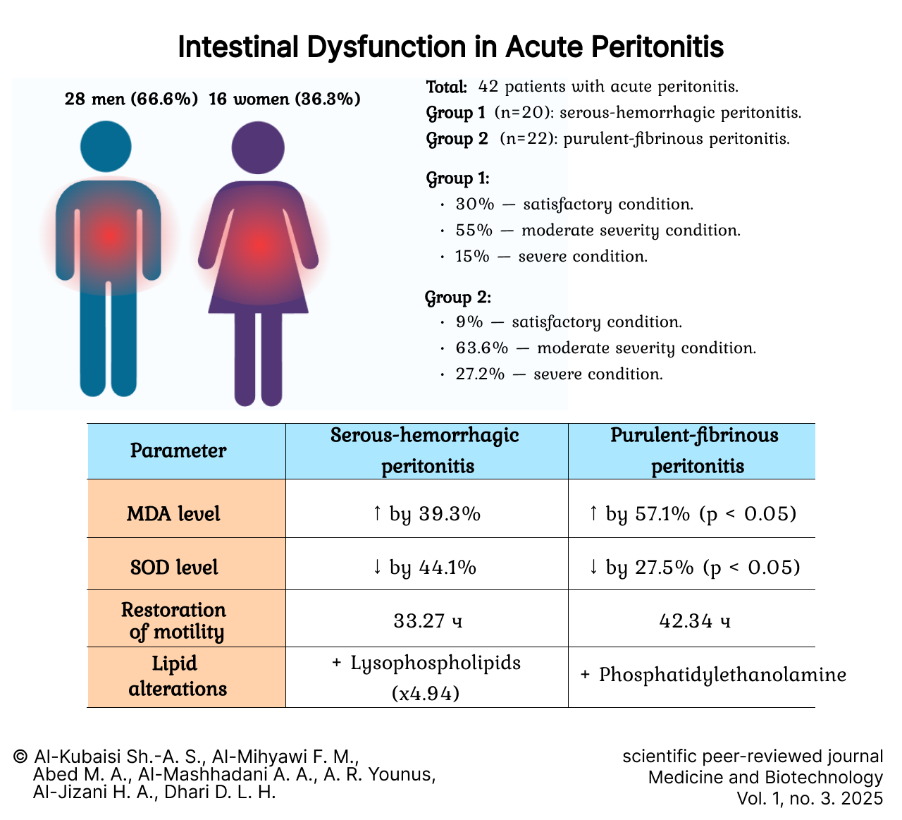
The clinical section includes 42 patients with acute peritonitis from whom informed consent was obtained. In the first group (20 patients), the morphofunctional state of the intestines in acute serous-hemorrhagic peritonitis was studied. In the second group (22 patients), the study was conducted on acute purulent-fibrinous peritonitis.
The average age was 51.67 (±5.27) years, there were 28 (66.7%) men and 14 (33.3%) women.
The morphofunctional state of the intestine in patients with peritonitis, as well as in the experiment, was assessed by blood supply and bio-energy of the organ tissues.
Due to the specifics of conducting research in the clinic, it was possible to study these parameters only during surgery.
The diseases that led to the development of peritonitis in patients were acute intestinal obstruction, acute appendicitis, perforated stomach ulcer, abdominal trauma.
The following methods were used in the work: determination of the oxidizing recovery potential (ORP), venous gradient by the Landis method, oxygen diffusion coefficient (ODC) in tissues, blood filling of small intestine tissues, extraction of lipids from small intestine tissues, the content of diene conjugates and malondialdehyde and superoxide dismutase activity.
Statistical processing of these results was performed using Excel 7.0 and Statistica 7.0 programs.
RESULTS
According to the prevalence of peritoneal lesions, peritonitis was diffuse in 14 (70.0%) of the first group and 16 (72.7%) of the second group, and localized in 6 (30.0%) and 6 (27.2%), respectively.
In patients of the first group, the disease was mainly reactive, in the second – toxic stage.
Analyzing patients by the duration of the disease, it was found that the severity of peritonitis in both groups depended on the duration of the disease that caused peritonitis.
The general condition of patients with acute peritonitis at admission to the surgical clinic was different. Satisfactory condition was determined in 6 (30.0%) patients of the first group and 2 (9.0%) of the second group, moderate severity – in 11 (55.0%) and 14 (63.6%), severe – in 3 (15.0%) and 6 (27.2%).
When studying the state of some bioenergetic processes in the intestine, it was found that in acute serous peritonitis, the electrogenesis of intestinal tissue structures is disrupted: the redox potential decreased by 28.4% (p < 0.05), the oxygen diffusion coefficient by 57.3% (p < 0.05). Blood supply also suffered: it increased by 82.1% (p < 0.05).
When analyzing similar indicators for purulent-fibrinous peritonitis, their large deviations from normal values were revealed. Significant changes were noted in blood filling parameters and oxygen diffusion coefficient. Thus, the deviation of the first indicator between the groups was 21.7% (p < 0.05), the second – 38.2% (p < 0.05) (fig. 1).

Fig.1. Blood supply and bioenergetics of the small intestine in patients
Note: here and further * – the significance of the difference to the norm at p < 0.05,
*1 – the significance of the difference in relation to the data of the first group at p < 0.05,
ORP – oxidizing recovery potential, ODC – oxygen diffusion coefficient
Source: the authors create all the figures
The analysis of the biopsy material revealed significant changes in the qualitative and quantitative composition of lipids in the tissue structures of the small intestine in acute serous peritonitis.
We have identified quite pronounced changes in the composition of phospholipids. The analysis of the fractional composition shows that significant deviations were detected in such labile fractions as lysophospholipids and phosphatidylcholine: the level of the former increased by 4.94 times (p < 0.01), the latter decreased by 27.5% (p < 0.05) (fig. 2).

Fig. 2. Lipid composition in small intestinal tissue in patients
An analysis of the literature data revealed that endogenous intoxication plays a major role in the pathogenetic mechanisms of acute peritonitis. It can progress the inflammatory response, enhance pathogenetic changes in tissues and organs, such as hypoxia, microcirculation, etc. This in turn leads to cellular changes. It has been shown that in acute peritonitis, changes in lipid metabolism are observed, which are associated with an intense inflammatory process and a systemic reaction of the body [5]. When analyzing the state of the key mechanisms regulating the aggregate state of tissue structures – the processes of lipid peroxidation, the state of the antioxidant system, it was revealed that they also depend on the form of acute peritonitis [6].
In acute purulent-fibrinous peritonitis in the tissue structures of the small intestine, changes in the qualitative and quantitative composition of lipids were more pronounced.
The most noticeable changes were in the levels of free fatty acids, total phospholipids, and cholesterol. Thus, compared with the first group of patients, the content of total fatty acids in intestinal tissue structures was higher by 26.7% (p < 0.05), total phospholipids and cholesterol were lower by 14.8% and 21.8%, respectively (p < 0.05). In other fractions studied, changes were noted in comparison with the norm. However, there were no significant differences compared to the first group.
The composition of phospholipids in acute purulent-fibrinous peritonitis in the tissue structures of the small intestine was also modified to a large extent.
It should be emphasized that, in general, changes in the qualitative and quantitative composition of phospholipids in the more severe form of peritonitis were also more pronounced. Other things are also noted. The most noticeable changes were in the levels of lysophospholipids, phosphatidylcholine, and phosphatidylethanolamine. Thus, compared with the first group, the levels of lysophospholipids and phosphatidylethanolamine increased by 29.3 and 20.7% respectively (p < 0.05), phosphatidylcholine decreased by 17.8% (p < 0.05).
It turned out that in acute serous peritonitis, free radical processes are activated in the tissues of the small intestine. An increase in the level of primary and secondary molecular products of lipid peroxidation was revealed. These changes were accompanied by a decrease in the antioxidant potential of organ tissues, which was recorded by a decrease (by 44.1%) in the activity of the key antioxidant enzyme, superoxide dismutase (fig. 3).

Fig. 3. Activity of lipoperoxidation and antioxidant systems in patients
Note: DC – diene conjugates, MDA – malonic dialdehyde, SOD – superoxide dismutase
Clinical studies have established that in acute purulent-fibrinous peritonitis, the processes of lipid peroxidation in the tissues of the small intestine become more pronounced. At the same time, the enzymatic antioxidant potential decreases to a greater extent compared with the first group.
Thus, the level of primary products of lipid peroxidation of DC increased by 39.3% (p < 0.05), respectively, compared with the first group, and the activity of superoxide dismutase decreased by 27.5% (p < 0.05).
It should also be noted that in acute purulent-fibrinous peritonitis, intestinal motility was restored after 42.34 (±1.15) h, whereas in acute serous peritonitis it was restored after 33.27 (±1.34) h (p < 0.05).
DISCUSSION AND CONCLUSION
Thus, the analysis of the obtained clinical data on the assessment of the morphofunctional state of the intestine in patients with peritonitis shows that the severity of changes on the part of the organ depends on the severity of peritonitis. Of course, the most important fact is the discovered fact that the severity of changes on the part of the intestine depends on membrane-destabilizing processes. The reason for this is the information found on changes in lipid metabolism, especially the lipid bilayer of cellular structures.
Clinical results also show that membrane-destructive phenomena in the organ are accompanied by activation of lipid peroxidation processes and a decrease in antioxidant enzyme potential.
About the authors
Shekh-Ahmed Saad Al-Kubaisi
National Research Mordovia State University
Author for correspondence.
Email: shekhahmed88@yandex.ru
ORCID iD: 0000-0003-4984-2674
Cand.Sci. (Med.), Associate Professor of the Department of Faculty Surgery
Russian Federation, 68 Bolshevistskaya St., Saransk 430005Farooq Mhammed Al-Mihyawi
National Research Mordovia State University
Email: Fq0000@bk.ru
ORCID iD: 0009-0002-5620-6895
Undergraduate Student, Medical Institute
Russian Federation, 68 Bolshevistskaya St., Saransk 430005Mohammed Ali Abed
National Research Mordovia State University
Email: moh0770moh00@gmail.com
ORCID iD: 0009-0005-6264-1684
Undergraduate Student, Medical Institute
Russian Federation, 68 Bolshevistskaya St., Saransk 430005Ahmed Ali Al-Mashhadani
National Research Mordovia State University
Email: ahmedalmshhdani1999@gmail.com
ORCID iD: 0009-0000-8794-8109
Undergraduate Student, Medical Institute
Russian Federation, 68 Bolshevistskaya St., Saransk 430005Asmaa Rabeea Younus
National Research Mordovia State University
Email: asmaarabea725@gmail.com
ORCID iD: 0009-0007-5525-3266
Undergraduate Student, Medical Institute
Russian Federation, 68 Bolshevistskaya St., Saransk 430005Hayder Abdulwahid Al-Jizani
National Research Mordovia State University
Email: hayderalhassany08@gmail.com
ORCID iD: 0009-0006-7690-2395
Undergraduate Student, Medical Institute
Russian Federation, 68 Bolshevistskaya St., Saransk 430005Layth Hasan Dhari Dhari
National Research Mordovia State University
Email: 3c76vclg@gmail.com
ORCID iD: 0009-0003-2979-8984
Undergraduate Student, Medical Institute
Russian Federation, 68 Bolshevistskaya St., Saransk 430005References
- Kumar D., Garg I., Sarwar A.H., Kumar L., Kumar V., Ramrakhia S. et al. Causes of Acute Peritonitis and Its Complication. Cureus. 2021;13(5):e15301. https://doi.org/10.7759/cureus.15301
- Fallani G., Lombardi R., Masetti M., Chisari M., Zanini N., Cattaneo G.M. et al. Urgent and Emergency Surgery for Secondary Peritonitis during the COVID-19 Outbreak: an Unseen Burden of a Healthcare Crisis. Updates in Surgery. 2021;73(2):753–762. https://doi.org/10.1007/s13304-020-00943-y
- Sattarov Sh.Kh., Ruzibaev S.A., Khursanov Y.E. Optimization of the Way of Correction of Endotoxicosis in Acute Peritonitis (Literature Review). Research Focus. 2022;1(2):144–150. (In Russ.). https://doi.org/10.5281/zenodo.7324431
- Lucas R., Hadizamani Y., Gonzales J., Gorshkov B., Bodmer T., Berthiaume Y. et al. Impact of Bacterial Toxins in the Lungs. Toxins (Basel). 2020;12(4):223. https://doi.org/10.3390/toxins12040223
- Al-Kubaisi Sh.A.S., Vlasov A.P., Myshkina N.A., Kumaksheva T.N., Hosina E.A., Romanov D.A. et al. Hemostatic Disorders in Acute Progressive Peritonitis. University Proceedings. Volga Region. Medical Sciences. 2023;1(65):14–24. (In Russ.). https://doi.org/10.21685/2072-3032-2023-1-2
- Vlasov A.P., Markin O.V., Vlasova T.I., Hosina E.A., Kumaksheva T.N., Myshkina N.A. et al. Liver Damage in Acute Peritonitis. Infekcii v hirurgii = Infections in Surgery. 2022;20(2):78–82. (In Russ.). https://elibrary.ru/pbwbho
Supplementary files

