

")


Potential use of radiomics analysis of cine-mode cardiac MRI to detect post-infarction lesions in the left ventricular myocardium
- Авторлар: Maksimova A.S.1, Samatov D.S.2, Merzlikin B.S.2, Shelkovnikova T.A.1, Listratov A.I.3, Zavadovsky K.V.1
-
Мекемелер:
- Tomsk National Research Medical Center of the Russian Academy of Sciences
- National Research Tomsk Polytechnic University
- Siberian State Medical University
- Шығарылым: Том 5, № 4 (2024)
- Беттер: 682-694
- Бөлім: Original Study Articles
- URL: https://bakhtiniada.ru/DD/article/view/309829
- DOI: https://doi.org/10.17816/DD630602
- ID: 309829
Дәйексөз келтіру

Аннотация
BACKGROUND: The size and location of an infarct lesion and its clear differentiation from normal tissue are important for clinical diagnosis and precision medicine. This paper is based on the study of radiomic attributes for differentiation of infarct and non infarct tissue using non contrast enhanced cine mode cardiac magnetic resonance imaging (MRI) data.
AIM: The aim of the study was to evaluate the potential use and informative value of radiomics analysis to identify post-infarction lesions in the left ventricular myocardium in patients with ischemic cardiomyopathy (ICM) using non-contrast-enhanced cine-mode cardiac MRI.
MATERIALS AND METHODS: Results of contrast-enhanced cardiac MRI were evaluated in 33 patients following surgical treatment for ICM. Texture analysis was performed on 66 lesions in cine-mode cardiac MRI images, and 105 texture attributes were determined for each lesion. Cardiac MRI was performed according to a standard technique using a Vantage Titan 1.5 T MRI scanner (Toshiba). For texture analysis, 3D Slicer version 5.2.2 (Pyradiomics) was used.
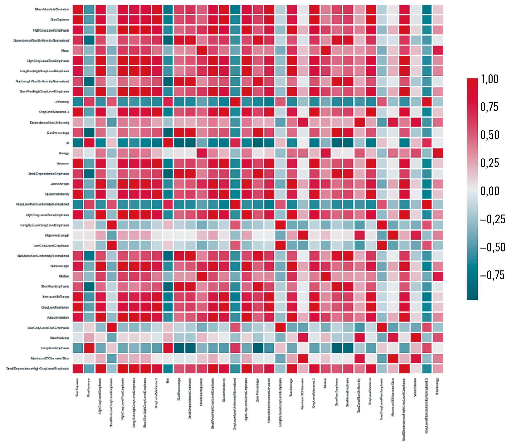
RESULTS: During the study, attribute collinearity diagrams were plotted, zero-significance attributes were identified, and attribute significance was determined using a gradient boosting algorithm, and the cumulative significance of attributes was estimated as a function of their total number. By identifying low-significance attributes, the least significant parameters that did not affect the overall significance level were determined. When single-valued attributes were extracted, no corresponding attributes were found. Based on the analysis results, an ROC curve was constructed for Lasso logistic regression (Se=57.14%, Sp=71.43%, AUC=0.76). The main result of this study was to determine radiomic attributes that characterized lesions corresponding to post-infarction cardiosclerosis and intact left ventricular wall based on cine-mode cardiac MRI images.
CONCLUSIONS: This study demonstrated that radiomics analysis of non-contrast-enhanced cine-mode cardiac MRI images is a promising approach to identify lesions corresponding to myocardial infarction and intact wall. This method may potentially be used to identify lesions of post-infarction cardiosclerosis in patients with ICM without contrast enhancement.
Толық мәтін
##article.viewOnOriginalSite##Авторлар туралы
Aleksandra Maksimova
Tomsk National Research Medical Center of the Russian Academy of Sciences
Хат алмасуға жауапты Автор.
Email: asmaximova@yandex.ru
ORCID iD: 0000-0002-4871-3283
SPIN-код: 2879-9550
MD, Cand. Sci. (Medicine)
Ресей, TomskDenis Samatov
National Research Tomsk Polytechnic University
Email: denissamatov470@gmail.com
ORCID iD: 0009-0000-1821-323X
Ресей, Tomsk
Boris Merzlikin
National Research Tomsk Polytechnic University
Email: merzlikin@tpu.ru
ORCID iD: 0000-0001-8545-9491
SPIN-код: 4815-6169
Cand. Sci. (Physics and Mathematics)
Ресей, TomskTatiana Shelkovnikova
Tomsk National Research Medical Center of the Russian Academy of Sciences
Email: fflly@mail.ru
ORCID iD: 0000-0001-8089-2851
SPIN-код: 1826-7850
MD, Cand. Sci. (Medicine)
Ресей, TomskArtem Listratov
Siberian State Medical University
Email: listrat312@gmail.com
ORCID iD: 0009-0004-3202-8179
Ресей, Tomsk
Konstantin Zavadovsky
Tomsk National Research Medical Center of the Russian Academy of Sciences
Email: Konstz@cardio-tomsk.ru
ORCID iD: 0000-0002-1513-8614
SPIN-код: 5081-3495
MD, Dr. Sci. (Medicine)
Ресей, TomskӘдебиет тізімі
- Shalnova SA, Drapkina OM, Kutsenko VA, et al. Myocardial infarction in the population of some Russian regions and its prognostic value. Russian Journal of Cardiology. 2022;27(6):4952. EDN: OCPROJ doi: 10.15829/1560-4071-2022-4952
- Desai R, Mishra V, Chhina AK, et al. Cardiovascular disease risk factors and outcomes of acute myocardial infarction in young adults: evidence from 2 nationwide cohorts in the United States a decade apart. Curr Probl Cardiol. 2023;48(9):101747. doi: 10.1016/j.cpcardiol.2023.101747
- Martins Marques T, Hausenloy DJ, Sluijter JP, et al. Girao Intercellular communication in the heart: therapeutic opportunities for cardiac ischemia. Trends Mol Med. 2021;27:248–262. doi: 10.1016/j.molmed.2020.10.002
- Schuleri KH, Centola M, Evers KS, et al. Cardiovascular magnetic resonance characterization of peri-infarct zone remodeling following myocardial infarction. J Cardiovasc Magn Reson. 2012;14:24. doi: 10.1186/1532-429X-14-24
- Bodi V, Monmeneu JV, Ortiz Perez JT, et al. Prediction of Reverse Remodeling at Cardiac MR Imaging Soon after First ST-Segment-Elevation Myocardial Infarction: Results of a Large Prospective Registry. Radiology. 2016;278:54–63. doi: 10.1148/radiol.2015142674
- Del Buono MG, Garmendia CM, Seropian IM, et al. Heart Failure After ST-Elevation Myocardial Infarction: Beyond Left Ventricular Adverse Remodeling. Curr Probl Cardiol. 2022;48(8):101215. doi: 10.1016/j.cpcardiol.2022.101215
- Ibanez B, Aletras AH, Arai AE, et al. Cardiac MRI Endpoints in Myocardial Infarction Experimental and Clinical Trials: JACC Scientific Expert Panel. J Am Coll Cardiol. 2019;74(2):238–256. doi: 10.1016/j.jacc.2019.05.024
- Ussov WYu, Babokin VE, Mochula OV, et al. Contrast-enhanced magnetic resonance tomography in patients with myocardial infarction and supraventricular tachyarrhythmias. Siberian Journal of Clinical and Experimental Medicine. 2014;29(4):33–38. EDN: TBFGPX doi: 10.29001/2073-8552-2014-29-4-33-38
- Usov VYu, Vyshlov EV, Mochula OV, et al. Contrast-ehanced MRI in time structure analysis of myocardial damage in acute infarction and early prehospital thrombolytic therapy. Medical Visualization. 2018;(2):56–69. EDN: XMLLXN doi: 10.24835/1607-0763-2018-2-56-69
- Kuo PH, Kanal E, Abu-Alfa AK, Cowper SE Gadolinium-based MR contrast agents and nephrogenic systemic fibrosis. Radiology. 2007;242(3):647–649. doi: 10.1148/radiol.2423061640
- Kim RJ, Wu E, Rafael A, et al. The use of contrast enhanced magnetic resonance imaging to identify reversible myocardial dysfunction. N Engl J Med. 2000;343(20):1445–1453. doi: 10.1056/NEJM200011163432003
- Kotu LP, Engan K, Eftestol T, et al. Segmentation of scarred and non scarred myocardium in LG enhanced CMR images using intensity based textural analysis. Annu Int Conf IEEE Eng Med Biol Soc. 2011:5698–5701. doi: 10.1109/IEMBS.2011.6091379
- Larroza A, Lopez Lereu MP, Monmeneu JV, et al. Texture analysis of cardiac cine magnetic resonance imaging to detect nonviable segments in patients with chronic myocardial infarction. Med Phys. 2018;45(4):1471–1480. doi: 10.1002/mp.12783
- Maksimova AS, Ussov WYu, Shelkovnikova TA, et al. Cardiac MRI Radiomics: review. Siberian Journal of Clinical and Experimental Medicine. 2023;38(3):13–22. EDN: RUADYI doi: 10.29001/2073-8552-2023-39-3-13-22
- Larroza A, Materka A, Lopez Lereu MP, et al. Differentiation between acute and chronic myocardial infarction by means of texture analysis of late gadolinium enhancement and cine cardiac magnetic resonance imaging. Eur J Radiol. 2017;92:78–83. doi: 10.1016/j.ejrad.2017.04.024
- Avard E, Shiri I, Hajianfar G, et al. Non contrast Cine Cardiac Magnetic Resonance image radiomics features and machine learning algorithms for myocardial infarction detection. Comput Biol Med. 2022;141:105145. doi: 10.1016/j.compbiomed
- Felker GM, Shaw LK, O’Connor CM A standardized definition of ischemic cardiomyopathy for use in clinical research. J Am Coll Cardiol. 2002;39(2):210–218. doi: 10.1016/s0735-1097(01)01738-7
- Liu M, Xin A, Chen T, et al. Non contrast cine cardiac magnetic resonance derived radiomics for the prediction of left ventricular adverse remodeling in patients with ST-segment elevation myocardial infarction. Korean J Radiol. 2023;24(9):827–837. doi: 10.3348/kjr.2023.0061
- Ma Q, Ma Y, Yu T, et al. Radiomics of non contrast enhanced T1 mapping: diagnostic and predictive performance for myocardial injury in acute ST-segment-elevation myocardial infarction. Korean J Radiol. 2021;22(4):535–46. doi: 10.3348/kjr.2019.0969
- Ma Q, Ma Y, Wang X, et al. A radiomic nomogram for prediction of major adverse cardiac events in ST-segment elevation myocardial infarction. Eur Radiol. 2021;31(2):1140–1150. doi: 10.1007/s00330-020-07176-y
- Chen BH, An DA, He J, et al. Myocardial extracellular volume fraction radiomics analysis for differentiation of reversible versus irreversible myocardial damage and prediction of left ventricular adverse remodeling after ST-elevation myocardial infarction. Eur Radiol. 2021;31(1):504–514. doi: 10.1007/s00330-020-07117-9
- Chang S, Han K, Kwon Y, et al. T1 Map-based radiomics for prediction of left ventricular reverse remodeling in patients with non ischemic dilated cardiomyopathy. Korean J Radiol. 2023;24:395–405. doi: 10.3348/kjr.2023.0065
- Frederiksen H, Iorgoveanu C, Mahi A. State of the Art and New Advances: Cardiac MRI. New Advances in Magnetic Resonance Imaging. 2023. Available from: http://dx.doi.org/10.5772/intechopen.112413. doi: 10.5772/intechopen.112413
- Bodi V, Monmeneu JV, Ortiz Perez JT, et al. Prediction of Reverse Remodeling at Cardiac MR Imaging Soon after First ST-Segment Elevation Myocardial Infarction: Results of a Large Prospective Registry. Radiology. 2016;278(1):54–63. doi: 10.1148/radiol.2015142674
Қосымша файлдар


