

")


人工智能程序在黄斑疾病诊断中的可行性研究
- 作者: Khabazova M.R.1, Ponomareva E.N.1, Loskutov I.A.2, Katalevskaya E.А.3, Sizov A.Y.3,4, Gabaraev G.М.1
-
隶属关系:
- Federal Research and Clinical Center of Specialized Medical Care and Medical Technologies
- Moscow Regional Research and Clinical Institute
- Digital Vision Solutions LLC
- Nizhny Novgorod State Technical University n.a. R.E. Alekseev
- 期: 卷 5, 编号 1 (2024)
- 页面: 17-28
- 栏目: 原创性科研成果
- URL: https://bakhtiniada.ru/DD/article/view/262945
- DOI: https://doi.org/10.17816/DD624131
- ID: 262945
如何引用文章

详细
论证。黄斑疾病是一大类病症。它们会导致视力丧失和视力低下。对这些病变的早期诊断对治疗策略的选择起着重要作用,它是疗效预测的决定性因素之一。
目的。本研究的目的是研究人工智能程序在基于对结构光学相干断层扫描图片的分析诊断黄斑疾病方面的可行性。
材料与方法。本研究对象包括在俄罗斯联邦医疗和生物局联邦专业医疗救护和医疗技术科学与临床中心以及以M.F.弗拉基米尔斯基莫斯科州临床研究所接受检查和治疗的患者。对200只有黄斑病变的眼和无黄斑病变的眼进行了检查。对RTVue XR 110-2眼科断层扫描仪上的结构光学相干断层扫描进行了临床对比分析。利用Retina.AI软件对光学相干断层扫描进行分析。
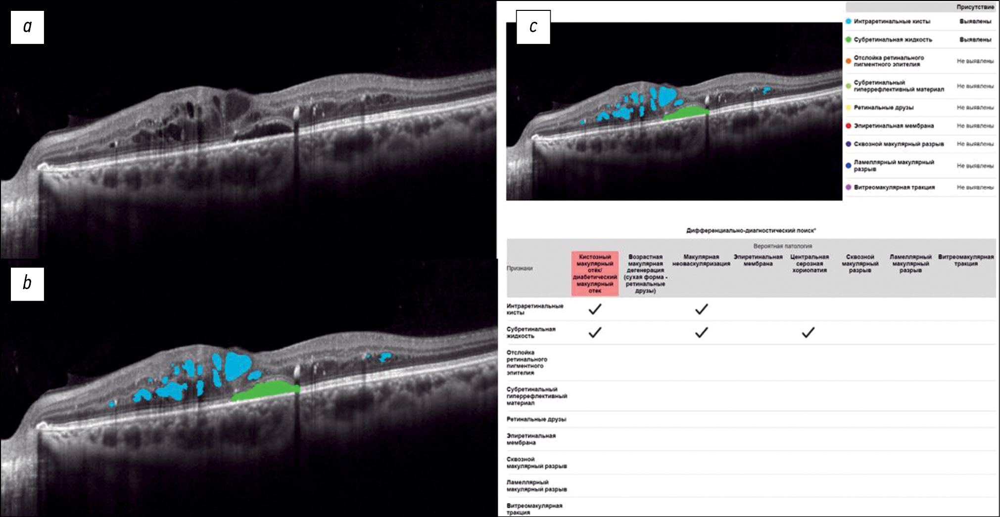
结果。使用该程序分析光学相干断层扫描图片时,确定了黄斑区的各种病理结构。此外,还得出了关于可能病理的结论。对获得的结果与眼科医生的结论进行了比较。该方法的灵敏度为95.16%;特异性为97.76%;准确率为97.38%。
结论。Retina.AI平台使眼科医生能够成功地对结构光学相干断层扫描图片进行自动分析,并检测眼底的各种病理状态。
作者简介
Margarita R. Khabazova
Federal Research and Clinical Center of Specialized Medical Care and Medical Technologies
Email: rita.khabazova@mail.ru
ORCID iD: 0000-0002-7770-575X
SPIN 代码: 2736-9089
俄罗斯联邦, Moscow
Elena N. Ponomareva
Federal Research and Clinical Center of Specialized Medical Care and Medical Technologies
Email: ponomareva.en@fnkc-fmba.ru
ORCID iD: 0009-0001-0828-9844
SPIN 代码: 7868-4425
俄罗斯联邦, Moscow
Igor A. Loskutov
Moscow Regional Research and Clinical Institute
Email: loskoutigor@mail.ru
ORCID iD: 0000-0003-0057-3338
SPIN 代码: 5845-6058
MD, Dr. Sci. (Medicine)
俄罗斯联邦, MoscowEvgenia А. Katalevskaya
Digital Vision Solutions LLC
Email: ekatalevskaya@mail.ru
ORCID iD: 0000-0002-5710-9205
SPIN 代码: 7849-8890
MD, Cand. Sci. (Medicine)
俄罗斯联邦, MoscowAlexander Yu. Sizov
Digital Vision Solutions LLC; Nizhny Novgorod State Technical University n.a. R.E. Alekseev
Email: sizov_ost_vk@mail.ru
ORCID iD: 0000-0003-3338-4015
SPIN 代码: 4468-1730
俄罗斯联邦, Moscow; Nizhny Novgorod
Georgiy М. Gabaraev
Federal Research and Clinical Center of Specialized Medical Care and Medical Technologies
编辑信件的主要联系方式.
Email: geor_gabaraev1@mail.ru
ORCID iD: 0000-0002-0759-3107
SPIN 代码: 1802-3224
俄罗斯联邦, Moscow
参考
- Report of the 2030 targets on effective coverage of eye care [Internet]. Geneva: World Health Organization. c2024. [cited 2023 Jan 1]. Available from: https://www.who.int/publications/i/item/9789240058002
- GBD 2019 Blindness and Vision Impairment Collaborators. Causes of blindness and vision impairment in 2020 and trends over 30 years, and prevalence of avoidable blindness in relation to VISION 2020: the Right to Sight: an analysis for the Global Burden of Disease Study. Lancet Glob Health. 2021;9(2):144–160. doi: 10.1016/S2214-109X(20)30489-7
- Samanta A, Aziz AA, Jhingan M, et al. Emerging Therapies in Neovascular Age-Related Macular Degeneration in 2020. Asia Pac J Ophthalmol (Phila). 2020;9(3):250–259. doi: 10.1097/APO.0000000000000291
- Stahl A. The Diagnosis and Treatment of Age-Related Macular Degeneration. Dtsch Arztebl Int. 2020;117:513–520. doi: 10.3238/arztebl.2020.0513
- Teo ZL, Tham YC, Yu M, et al. Global Prevalence of Diabetic Retinopathy and Projection of Burden through 2045: Systematic Review and Meta-analysis. Ophthalmology. 2021;128(11):1580–1591. doi: 10.1016/j.ophtha.2021.04.027
- Schaal S, Kaplan HJ, editors. Cystoid Macular Edema. Switzerland: Springer International Publishing; 2017. doi: 10.1007/978-3-319-39766-5
- Bikbov MM, Fayzrakhmanov RR, Zaynullin RM, et al. Macular oedema as manifestation of diabetic retinopathy. Diabetes mellitus. 2017;20(4):263–269. EDN: ZMZAON doi: 10.14341/DM8328
- Chernykh DV, Chernykh VV, Trunov AN. Cytokines and growth factors in the pathogenesis of proliferative diabetic retinopathy. Moscow: Oftal’mologiya; 2017. EDN: ZNDEWH
- Gupta A, Tripathy K. Central Serous Chorioretinopathy [Internet]. [Updated 2022 Aug 22]. In: StatPearls [Internet]. Treasure Island (FL): StatPearls Publishing, 2022. Available from: https://www.statpearls.com/point-of-care/96027
- Semeraro F, Morescalchi F, Russo A, et al. Central Serous Chorioretinopathy: Pathogenesis and Management. Clinical ophthalmology. 2019;13:2341–2352. doi: 10.2147/OPTH.S220845
- Oh KT, Lazzaro DR, editors. Macular Hole. [Internet]. Medscape, 2020. [cited 2020 Jan 02]. Available from: https://emedicine.medscape.com/article/1224320-overview#a6
- Darian-Smith E, Howie AR, Allen PL, et al. Tasmanian macular hole study: whole population-based incidence of full thickness macular hole. Clinical & Experimental Ophthalmology. 2016;44(9):812–816. doi: 10.1111/ceo.12801
- Fung AT, Galvin J, Tran T. Epiretinal membrane: A review. Clinical & Experimental Ophthalmology. 2021;49:289–308. doi: 10.1111/ceo.13914
- Oh KT, Lazzaro DR, editors. Epiretinal Membrane [Internet]. Medscape, 2020. [cited 2020 Jan 02]. Available from: https://emedicine.medscape.com/article/1223882-overview#a4
- World Health Organization. Regional Office for Europe. Screening for diabetic retinopathy: a short guide. Increase effectiveness, maximize benefits and minimize harm [Internet]. Copenhagen; 2021. [cited 2020 Jan 02]. Available from: https://www.who.int/europe/publications/i/item/9789289055321
- Qassimi AN, Kozak I, Karam AM, et al. Management of Diabetic Macular Edema: Guidelines from the Emirates Society of Ophthalmology. Ophthalmology and therapy. 2022;11:1937–1950. doi: 10.1007/s40123-022-00547-2
- Katalevskaya EA, Katalevskiy DYu, Tyurikov MI, Velieva IA, Bol’shunov AV. Future of artificial intelligence for the diagnosis and treatment of retinal diseases. Russian journal of clinical ophthalmology. 2022;22(1):36–43. EDN: AEBQGU doi: 10.32364/2311-7729-2022-22-1-36-43
- Schmidt-Erfurth U, Reiter GS, Riedl S, et al. AI-based monitoring of retinal fluid in disease activity and under therapy. Prog Retin Eye Res. 2022;86. doi: 10.1016/j.preteyeres.2021.100972
- Altris.ai [Internet]. United States: Altris Inc. [cited 2022 Jan 01]. Available from: https://www.altris.ai
- Malyugin BE, Sakhnov SN, Axenova LE, et al. A deep machine learning model development for the biomarkers of the anatomical and functional anti-VEGF therapy outcome detection on retinal OCT images. Fyodorov Journal of Ophthalmic Surgery. 2022;(S4):77–84. EDN: OWQLRM doi: 10.25276/0235-4160-2022-4S-77-84
补充文件


