

")


Ограничения использования гистологического исследования биоптатов как «золотого стандарта» диагностики на примере аденокарциномы пищевода: описание случая
- Авторы: Ахмедзянова Д.А.1, Юцевич О.К.2, Решетников Р.В.1, Тащян О.В.3, Пирогов С.С.2, Мазурова М.П.2, Волченко Н.Н.2, Камалов А.К.2, Шумская Ю.Ф.1, Мнацаканян М.Г.3
-
Учреждения:
- Научно-практический клинический центр диагностики и телемедицинских технологий
- Московский научно-исследовательский онкологический институт имени П.А. Герцена — филиал Национального медицинского исследовательского центра Радиологии
- Первый Московский государственный медицинский университет имени И.М. Сеченова
- Выпуск: Том 4, № 4 (2023)
- Страницы: 633-642
- Раздел: Клинические случаи и серии клинических случаев
- URL: https://bakhtiniada.ru/DD/article/view/262990
- DOI: https://doi.org/10.17816/DD561354
- ID: 262990
Цитировать

Аннотация
Аденокарцинома пищевода — одно из самых распространённых злокачественных новообразований желудочно-кишечного тракта. Для выявления заболевания на ранних стадиях используют эндоскопические, морфологические, иммуногистохимические методы обследования, однако они требуют не только применения высокоспециализированного оборудования, но и зависят от профессионализма врача-эндоскописта и врача-патоморфолога.
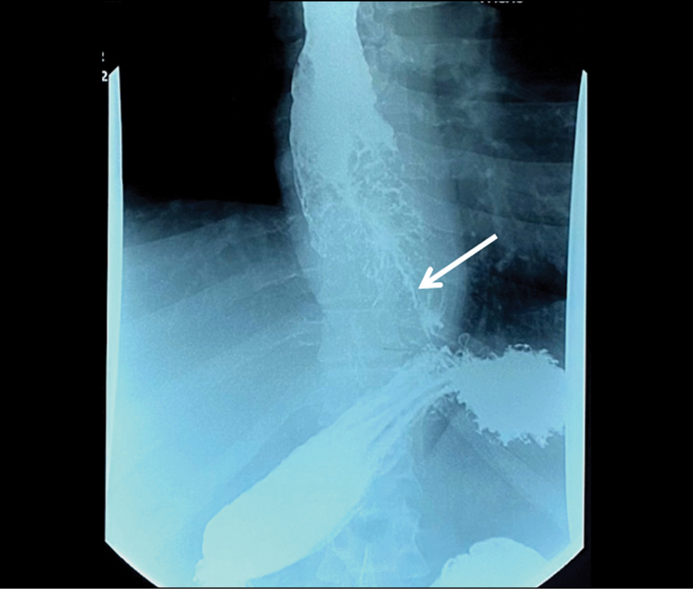
В статье описано клиническое наблюдение пациента с прогрессирующей дисфагией, вызванной опухолью пищевода, распространяющейся на субкардиальный отдел желудка, которую не удавалось патоморфологически верифицировать в течение одного года. Данные эзофагогастродуоденоскопии, выполненной в медицинской организации по месту жительства, компьютерной томографии и рентгеноскопии пищевода с двойным контрастированием не оставляли сомнений в злокачественности новообразования, однако результаты многочисленных гистологических исследований свидетельствовали в пользу аденомы пилорических желёз, аденомы из париетальных, либо онкоцитарных клеток с фокусами дисплазии эпителия высокой степени. Эндоскопическое исследование с таргетированной биопсией в условиях специализированного учреждения позволило доказать злокачественность опухолевого процесса.
Результаты исследования демонстрируют важность клинической картины и инструментальных методов для постановки заключительного диагноза при противоречивых данных патоморфологических исследований и в очередной раз поднимают проблему ограничений гистологического исследования биоптатов как «золотого стандарта» диагностики злокачественных новообразований.
Полный текст
Открыть статью на сайте журналаОб авторах
Дина Альфредовна Ахмедзянова
Научно-практический клинический центр диагностики и телемедицинских технологий
Автор, ответственный за переписку.
Email: AkhmedzyanovaDA@zdrav.mos.ru
ORCID iD: 0000-0001-7705-9754
SPIN-код: 6983-5991
Scopus Author ID: 58104960900
Россия, Москва
Ольга Константиновна Юцевич
Московский научно-исследовательский онкологический институт имени П.А. Герцена — филиал Национального медицинского исследовательского центра Радиологии
Email: o.yutsevitch@yandex.ru
ORCID iD: 0000-0002-3860-9853
Россия, Москва
Роман Владимирович Решетников
Научно-практический клинический центр диагностики и телемедицинских технологий
Email: r.reshetnikov@npcmr.ru
ORCID iD: 0000-0002-9661-0254
SPIN-код: 8592-0558
канд. ф.-м. наук
Россия, МоскваОльга Валерьевна Тащян
Первый Московский государственный медицинский университет имени И.М. Сеченова
Email: olgatash1@rambler.ru
ORCID iD: 0000-0001-6759-6820
SPIN-код: 3658-1120
канд. мед. наук
Россия, МоскваСергей Сергеевич Пирогов
Московский научно-исследовательский онкологический институт имени П.А. Герцена — филиал Национального медицинского исследовательского центра Радиологии
Email: pirogov@mail.ru
ORCID iD: 0000-0002-8101-2155
SPIN-код: 7812-5502
д-р мед. наук
Россия, МоскваМария Павловна Мазурова
Московский научно-исследовательский онкологический институт имени П.А. Герцена — филиал Национального медицинского исследовательского центра Радиологии
Email: mnioi_morphology@mail.ru
ORCID iD: 0000-0002-4873-4455
SPIN-код: 4455-3055
канд. мед. наук
Россия, МоскваНадежда Николаевна Волченко
Московский научно-исследовательский онкологический институт имени П.А. Герцена — филиал Национального медицинского исследовательского центра Радиологии
Email: mnioi_morphology@mail.ru
ORCID iD: 0000-0003-0421-4172
д-р мед. наук, профессор
Россия, МоскваАзиз Кураглиевич Камалов
Московский научно-исследовательский онкологический институт имени П.А. Герцена — филиал Национального медицинского исследовательского центра Радиологии
Email: kak6768@mail.ru
ORCID iD: 0000-0001-7376-6056
SPIN-код: 1671-1600
Россия, Москва
Юлия Федоровна Шумская
Научно-практический клинический центр диагностики и телемедицинских технологий
Email: ShumskayaYF@zdrav.mos.ru
ORCID iD: 0000-0002-8521-4045
SPIN-код: 3164-5518
Россия, Москва
Марина Генриковна Мнацаканян
Первый Московский государственный медицинский университет имени И.М. Сеченова
Email: mnatsakanyan08@mail.ru
ORCID iD: 0000-0001-9337-7453
SPIN-код: 2015-1822
д-р мед. наук, профессор
Россия, МоскваСписок литературы
- Bray F., Ferlay J., Soerjomataram I., et al. Global cancer statistics 2018: GLOBOCAN estimates of incidence and mortality worldwide for 36 cancers in 185 countries // CA Cancer J Clin. 2018. Vol. 68, N 6. P. 394–424. doi: 10.3322/caac.21492
- McColl K.E.L. What is causing the rising incidence of esophageal adenocarcinoma in the West and will it also happen in the East? // J Gastroenterol. 2019. Vol. 54, N 8. P. 669–673. doi: 10.1007/s00535-019-01593-7
- Joseph A., Raja S., Kamath S., et al. Esophageal adenocarcinoma: A dire need for early detection and treatment // Cleve Clin J Med. 2022. Vol. 89, N 5. P. 269–279. doi: 10.3949/ccjm.89a.21053
- Uhlenhopp D.J., Then E.O., Sunkara T., Gaduputi V. Epidemiology of esophageal cancer: update in global trends, etiology and risk factors // Clin J Gastroenterol. 2020. Vol. 13, N 6. P. 1010–1021. doi: 10.1007/s12328-020-01237-x
- Zhang H.Y., Spechler S.J., Souza R.F. Esophageal adenocarcinoma arising in Barrett esophagus // Cancer Lett. 2009. Vol. 275, N 2. P. 170–177. doi: 10.1016/j.canlet.2008.07.006
- Deng H.Y., Alai G., Luo J., et al. Cancerous esophageal stenosis before treatment was significantly correlated to poor prognosis of patients with esophageal cancer: a meta-analysis // J Thorac Dis. 2018. Vol. 10, N 7. P. 4212–4219. doi: 10.21037/jtd.2018.06.89
- Sillah K., Pritchard S.A., Watkins G.R., et al. The degree of circumferential tumour involvement as a prognostic factor in oesophageal cancer // Eur J Cardiothorac Surg. 2009. Vol. 36, N 2. P. 368–373. doi: 10.1016/j.ejcts.2008.12.052
- Deng H.Y., Li G., Luo J. Does oesophageal stenosis have any impact on survival of oesophageal cancer patients? // Interact Cardiovasc Thorac Surg. 2018. Vol. 27, N 3. P. 384–386. doi: 10.1093/icvts/ivy095
- Knight W.R.C., McEwen R., Byrne B.E., et al. Endoscopic tumour morphology impacts survival in adenocarcinoma of the oesophagus // Eur J Surg Oncol. 2020. Vol. 46, N 12. P. 2257–2261. doi: 10.1016/j.ejso.2020.07.003
- И–74 Информативность методов лучевой диагностики при различных патологических состояниях организма. Раздел 2. Диагностика патологических состояний и заболеваний желудочно-кишечного тракта / под ред. С.П. Морозова. Москва, 2018.
- Ishihara R., Goda K., Oyama T. Endoscopic diagnosis and treatment of esophageal adenocarcinoma: introduction of Japan Esophageal Society classification of Barrett’s esophagus // J Gastroenterol. 2019. Vol. 54, N 1. P. 1–9. doi: 10.1007/s00535-018-1491-x
- Загайнова Е.В., Загайнов В.Е., Гладкова Н.Д., и др. Оптическая когерентная томография при хирургическом лечении рака пищевода // Вестник хирургии имени И.И. Грекова. 2007. Т. 166(2. С. 22–26.
- Давыдов М.И., Тер-Ованесов М.Д., Стилиди И.С., и др. Пищевод Барретта: от теоретических основ к практическим рекомендациям // Практическая онкология. 2003. Т. 4, № 2. С. 109–119.
- Barber M.S., Aronson J.K., von Schoen-Angerer T., et al. Рекомендации CARE для описания случаев: разъяснения и уточнения // Digital Diagnostics. Vol. 3, N 1. C. 16–42. doi: 10.17816/DD105291
- Wani S., Rubenstein J.H., Vieth M., Bergman J. Diagnosis and Management of Low-Grade Dysplasia in Barrett’s Esophagus: Expert Review From the Clinical Practice Updates Committee of the American Gastroenterological Association // Gastroenterology. 2016. Vol. 151, N 5. P. 822–835. doi: 10.1053/j.gastro.2016.09.040
- di Pietro M., Canto M.I., Fitzgerald R.C. Endoscopic Management of Early Adenocarcinoma and Squamous Cell Carcinoma of the Esophagus: Screening, Diagnosis, and Therapy // Gastroenterology. 2018. Vol. 154, N 2. P. 421–436. doi: 10.1053/j.gastro.2017.07.041
- Winiker M., Mantziari S., Figueiredo S.G., et al. Accuracy of preoperative staging for a priori resectable esophageal cancer // Dis Esophagus. 2018. Vol. 31, N 1. P. 1–6. doi: 10.1093/dote/dox113
- Elsadek H.M., Radwan M.M. Diagnostic Accuracy of Mucosal Biopsy versus Endoscopic Mucosal Resection in Barrett’s Esophagus and Related Superficial Lesions // Int Sch Res Notices. 2015. Vol. 2015. doi: 10.1155/2015/735807
- Трякин А.А., Бесова Н.С., Волков Н.М., и др. Практические рекомендации по лекарственному лечению рака пищевода и пищеводно-желудочного перехода // Злокачественные опухоли. 2021. Т. 11, № 3S2-1. С. 299–313. doi: 10.18027/2224-5057-2021-11-3s2-20
- Ajani J.A., D’Amico T.A., Bentrem D.J., et al. Esophageal and Esophagogastric Junction Cancers, Version 2.2023, NCCN Clinical Practice Guidelines in Oncology // J Natl Compr Canc Netw. 2023. Vol. 21, N 4. P. 393–422. doi: 10.6004/jnccn.2023.0019
- Ormsby A.H., Petras R.E., Henricks W.H., et al. Observer variation in the diagnosis of superficial oesophageal adenocarcinoma // Gut. 2002. Vol. 51, N 5. P. 671–676. doi: 10.1136/gut.51.5.671
Дополнительные файлы


