The role of computed tomography in the differential diagnosis of an intracardiac mass of the mitral valve: a case series
- Authors: Onoyko M.V.1, Mershina E.A.1, Arakelyants A.A.1,2, Sinitsyn V.E.1
-
Affiliations:
- Lomonosov Moscow State University
- Sechenov First Moscow State Medical University
- Issue: Vol 5, No 4 (2024)
- Pages: 893-901
- Section: Case reports
- URL: https://bakhtiniada.ru/DD/article/view/309844
- DOI: https://doi.org/10.17816/DD629893
- ID: 309844
Cite item
Abstract
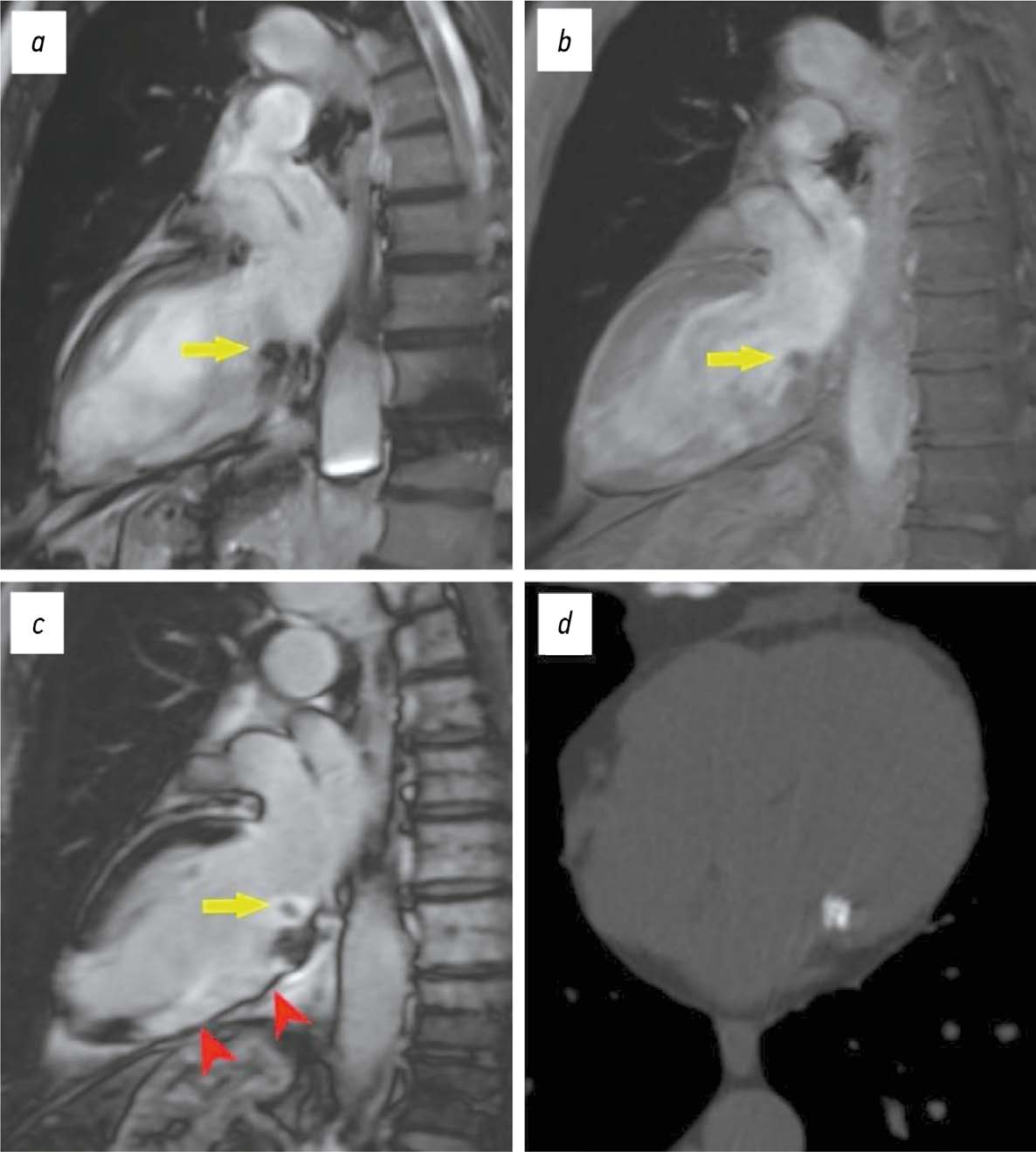
The differential diagnosis of an echocardiographically detected intracardiac mass in the mitral annulus can be challenging and usually requires a multimodal approach. This type of lesion is very often associated with subvalvular calcification of the mitral valve. The rare, caseous, variant is the most difficult to diagnose. This case series highlights the clinical significance of computed tomography in detecting and characterizing subvalvular mitral annular calcification when other modalities, particularly echocardiography, are inconclusive. The aim of this article was to raise awareness among specialists of the classic signs of caseous subvalvular calcification of the mitral annulus when visualized with different modalities. Special attention is also given to providing a differential diagnostic series that identifies features that differentiate subvalvular calcification of the mitral annulus from other conditions at this site. Healthcare professionals need to be aware of these mitral valve lesions in order to predict possible associated complications and plan a treatment strategy that may help avoid unnecessary surgical procedures in some cases.
Full Text
##article.viewOnOriginalSite##About the authors
Maria V. Onoyko
Lomonosov Moscow State University
Author for correspondence.
Email: onoykomary@gmail.com
ORCID iD: 0000-0002-7727-3360
SPIN-code: 6380-7495
MD
Russian Federation, MoscowElena A. Mershina
Lomonosov Moscow State University
Email: elena_mershina@mail.ru
ORCID iD: 0000-0002-1266-4926
SPIN-code: 6897-9641
MD, Cand. Sci. (Medicine), Assistant Professor
Russian Federation, MoscowAmalia A. Arakelyants
Lomonosov Moscow State University; Sechenov First Moscow State Medical University
Email: nxrrimma@mail.ru
ORCID iD: 0000-0002-1243-2471
SPIN-code: 4990-6008
MD, Cand. Sci. (Medicine)
Russian Federation, MoscowValentin E. Sinitsyn
Lomonosov Moscow State University
Email: vsini@mail.ru
ORCID iD: 0000-0002-5649-2193
SPIN-code: 8449-6590
MD, Dr. Sci. (Medicine), Professor
Russian Federation, MoscowReferences
- Savage DD, Garrison RJ, Castelli WP, et al. Prevalence of submitral (anular) calcium and its correlates in a general population based sample (the Framingham Study). Am J Cardiol. 1983;51(8):1375–1378. doi: 10.1016/0002-9149(83)90315-6
- Abramowitz Y, Jilaihawi H, Chakravarty T, et al. Mitral Annulus Calcification. J Am Coll Cardiol. 2015;66(17):1934–1941. doi: 10.1016/j.jacc.2015.08.872
- Kanjanauthai S, Nasir K, Katz R, et al. Relationships of mitral annular calcification to cardiovascular risk factors: the Multi Ethnic Study of Atherosclerosis (MESA). Atherosclerosis. 201;213(2):558–562. doi: 10.1016/j.atherosclerosis.2010.08.072
- Harpaz D, Auerbach I, Vered Z, et al. Caseous calcification of the mitral annulus: a neglected, unrecognized diagnosis. J Am Soc Echocardiogr. 2001;14(8):825–831. doi: 10.1067/mje.2001.111877
- Silbiger JJ. Anatomy, mechanics, and pathophysiology of the mitral annulus. Am Heart J. 2012;164(2):163–176. doi: 10.1016/j.ahj.2012.05.014
- Deluca G, Correale M, Ieva R, et al. The incidence and clinical course of caseous calcification of the mitral annulus: a prospective echocardiographic study. J Am Soc Echocardiogr. 2008;21(7):828–833. doi: 10.1016/j.echo.2007.12.004
- Akram M, Hasanin AM. Caseous mitral annular calcification: Is it a benign condition? J Saudi Hear Assoc. 2012;24(3):205–208. doi: 10.1016/j.jsha.2012.02.003
- Hamirani YS, Nasir K, Blumenthal RS, et al. Relation of mitral annular calcium and coronary calcium (from the Multi Ethnic Study of Atherosclerosis [MESA]). Am J Cardiol. 2011;107(9):1291–1294. doi: 10.1016/j.amjcard.2011.01.005
- Shriki J, Rongey C, Ghosh B, et al. Caseous mitral annular calcifications: Multimodality imaging characteristics. World J Radiol. 2010;2(4):143-147. doi: 10.4329/wjr.v2.i4.143
- Gravina M, Casavecchia G, Manuppelli V, et al. Mitral annular calcification: Can CMR be useful in identifying caseous necrosis? Interv Med Appl Sci. 2019;11(1):71-73. doi: 10.1556/1646.10.2018.47
- Mayr A, Müller S, Feuchtner G. The Spectrum of Caseous Mitral Annulus Calcifications. JACC Case Rep. 2021;3(1):104–108. doi: 10.1016/j.jaccas.2020.09.039
- Belkind MB, Butorova EA, Stukalova OV, et al. Caseous calcification of the mitral annulus. Eurasian heart journal. 2023;(4):90–93. doi: 10.38109/2225-1685-2023-4-90-93
- Saidova MA, Atabaeva LS, Stukalova OV. Caseous calcification of the mitral annulus. Russian Cardiology Bulletin. 2019;14(3):62–67. doi: 10.36396/MS.2019.14.03.010
Supplementary files


