

")


Multiple biliary microhamartomas diagnosed in an unsuspecting elderly patient
- Authors: Balbino M.1, Montatore M.1, Fascia G.1, Tupputi R.2, Masino F.1, Muscatella G.1, Mannatrizio D.1, Guglielmi G.1,2,3
-
Affiliations:
- University of Foggia
- Dimiccoli Hospital
- Casa Sollievo della Sofferenza Hospital
- Issue: Vol 5, No 2 (2024)
- Pages: 334-341
- Section: Case reports
- URL: https://bakhtiniada.ru/DD/article/view/264843
- DOI: https://doi.org/10.17816/DD623322
- ID: 264843
Cite item

Abstract
Multiple biliary hamartomas are a benign incidental finding in the liver. They are not easily detected if one has never seen them, and if appropriate imaging tests are unavailable, and also can be challenging to differentiate from other liver lesions based on imaging alone. Thus, this study aimed to expand the radiologist’s digital image library, enabling a quick and precise differential diagnosis. This paper also highlights the importance of thorough radiological assessment and need for a multidisciplinary approach, involving radiologists, hepatologists, and pathologists, to ensure a precise diagnosis.
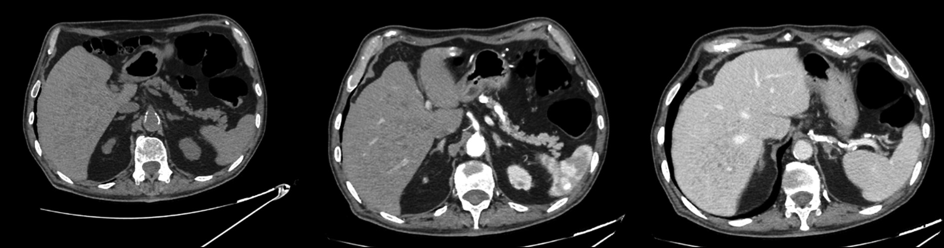
The patient presented at the hospital for a computed tomography scan and an abdominal magnetic resonance imaging recommended by his general practitioner to assess the biliary tree (magnetic resonance cholangiopancreatography), owing to persistent abdominal pain. The patient had never undergone an abdominal magnetic resonance imaging previously; hence, the discovery of hepatic lesions was incidental and unexpected.
Magnetic resonance imaging revealed multiple benign lesions in both the hepatic lobes comparable to the Von Meyenburg complex. These lesions are multiple hamartomas and behave differently in all magnetic resonance imaging sequences.
Images acquired with different magnetic resonance imaging sequences were carefully examined. Multiple lesions were found scattered throughout the liver; however, the lesions were benign and consistent with the diagnosis of multiple biliary hamartomas.
Medical practitioners should examine the presence of multiple biliary hamartomas and consider them in the differential diagnosis when patients present with hepatic abnormalities. This can prevent unnecessary interventions and guide appropriate patient management.
Full Text
##article.viewOnOriginalSite##About the authors
Marina Balbino
University of Foggia
Email: marinabalbino93@gmail.com
ORCID iD: 0009-0009-2808-5708
MD
Italy, FoggiaManuela Montatore
University of Foggia
Email: manuela.montatore@unifg.it
ORCID iD: 0009-0002-1526-5047
MD
Italy, FoggiaGiacomo Fascia
University of Foggia
Email: giacomo.fascia@unifg.it
ORCID iD: 0000-0001-5244-5093
MD
Italy, FoggiaRuggiero Tupputi
Dimiccoli Hospital
Email: rutudott@gmail.com
MD
Italy, BarlettaFederica Masino
University of Foggia
Email: federicamasino@gmail.com
MD
Italy, FoggiaGianmichele Muscatella
University of Foggia
Email: muscatella94@gmail.com
ORCID iD: 0009-0004-3535-5802
MD
Italy, FoggiaDomenico Mannatrizio
University of Foggia
Email: dr.mannatrizio@gmail.com
ORCID iD: 0000-0003-3365-7132
Italy, Foggia
Giuseppe Guglielmi
University of Foggia; Dimiccoli Hospital; Casa Sollievo della Sofferenza Hospital
Author for correspondence.
Email: giuseppe.guglielmi@unifg.it
ORCID iD: 0000-0002-4325-8330
MD, Professor
Italy, Foggia; Barletta; FoggiaReferences
- Zheng RQ, Zhang B, Kudo M, Onda H, Inoue T. Imaging findings of biliary hamartomas. World J Gastroenterol. 2005;11(40):6354–6359. doi: 10.3748/wjg.v11.i40.6354
- Gil-Bello D, Ballesteros E, Sanfeliu E, Andreu FJ. Calcification in biliary hamartomatosis. Br J Radiol. 2012;85(1012):e099–e101. doi: 10.1259/bjr/95019559
- Thommesen N. Biliary hamartomas (von Meyenburg complexes) in liver needle biopsies. Acta Pathol Microbiol Scand A. 1978;86(2):93–99. doi: 10.1111/j.1699-0463.1978.tb02019.x
- Aguado IC, Álvarez MH, Hernández JS, La Orden Izquierdo E. Hamartomatosis biliar en una lactante con colitis alérgica: revisión a propósito de un caso. Rev Pediatr Aten Primaria. 2013;15(59):e111–e114. doi: 10.4321/S1139-76322013000400014
- Horton KM, Bluemke DA, Hruban RH, Soyer P, Fishman EK. CT and MR imaging of benign hepatic and biliary tumors. Radiographics. 1999;19(2):431–451. doi: 10.1148/radiographics.19.2.g99mr0443
- Brancatelli G, Federle MP, Vilgrain V, et al. Fibropolycystic liver disease: CT and MR imaging findings. RadioGraphics. 2005;25(3):659–670. doi: 10.1148/rg.253045114
- Bravo-Acosta M, Rosendo-Namías J, Martínez-Méndez D. Hamartomatosis biliar múltiple: “imagen en cielo estrellado”. Rev Gastroenterol MEX. 2020;86(2). doi: 10.1016/j.rgmx.2020.08.002
- Choi BI, Yeon KM, Kim SH, et al. Caroli disease: central dot sign in CT. Radiology. 1990;174(1):161–163. doi: 10.1148/radiology.174.1.2294544
- Kin HK, Jin SY. Cholangiocarcinoma arising in von Meyenburg complexes. Korean J Hepatol. 2011;17(2):161–164. doi: 10.3350/kjhep.2011.17.2.161
- Song JS, Lee YJ, Kim KW, et al. Cholangiocarcinoma arising in von Meyenburg complexes: report of four cases. Pathol Int. 2008;58(8):503–512. doi: 10.1111/j.1440-1827.2008.02264.x
- Xu AM, Xian ZH, Zhang SH, Chen XF. Intrahepatic cholangiocarcinoma arising in multiple bile duct hamartomas: report of two cases and review of the literature. Eur J Gastroenterol Hepatol. 2009;21(5):580–584. doi: 10.1097/MEG.0b013e3282fc73b1
- Venkatanarasimha N, Thomas R, Armstrong EM, et al. Imaging features of ductal plate malformations in adults. Clin Radiol. 2011;66(11):1086–1093. doi: 10.1016/j.crad.2011.05.008
- Desmet VJ. Pathogenesis of ductal plate malformation. J Gastroenterol Hepatol. 2004;19(S7):S356–S360. doi: 10.1111/j.1440-1746.2004.03702.x
- Soreide K, Korner H, Havnen J, et al. Bile duct cysts in adults. Br J Surg. 2004;91(12):1538–1548. doi: 10.1002/bjs.4815
Supplementary files


