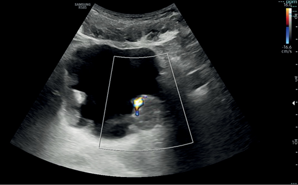
Conventional and innovative imaging modalities in bladder cancer: Techniques and applications
- Authors: Masino F.1, Eusebi L.2, Muscatella G.1, Montatore M.1, Sortino G.2, Giannubilo W.3, Guglielmi G.1,4,5
-
Affiliations:
- Foggia University School of Medicine
- Carlo Urbani Hospital
- Civitanova Marche Hospital
- Dimiccoli Hospital
- IRCCS Casa Sollievo della Sofferenza Hospital
- Issue: Vol 5, No 2 (2024)
- Pages: 318-333
- Section: Reviews
- URL: https://bakhtiniada.ru/DD/article/view/264841
- DOI: https://doi.org/10.17816/DD623889
- ID: 264841
Cite item
Abstract
This narrative review describes the current status of imaging in the evaluation of bladder cancer, considering conventional technologies such as ultrasonography, computed tomography urography, and magnetic resonance imaging, as well as novel technologies such as contrast-enhanced ultrasonography and dual-energy computed tomography.
The article is organized by first presenting an introduction on both the anatomy of the bladder (to understand its normal appearance on imaging) and the main features of bladder cancer with reference to epidemiology, clinical picture, classification, and treatment. Subsequently, the role of imaging is discussed, with an explanation of the technique and applications in bladder cancer assessment for each modality.
Imaging plays a critical role in the detection and staging of bladder cancer. In particular, the role of magnetic resonance imaging is expanding because it enables differentiating muscle-invasive bladder cancer from non-muscle-invasive bladder cancer using the Vesical Imaging-Reporting and Data System (VI-RADS), along with conventional technologies, such as computed tomography urography and ultrasonography. Contrast-enhanced ultrasound and dual-energy computed tomography are new imaging modalities that offer special advantages and provide the right approach to patients with oncological conditions. This review ends with the presentation of integrated imaging modalities such as positron emission tomography combined with computed tomography or magnetic resonance imaging, which are promising methods for bladder cancer staging.
Full Text
##article.viewOnOriginalSite##About the authors
Federica Masino
Foggia University School of Medicine
Email: federicamasino@gmail.com
ORCID iD: 0009-0004-4289-3289
MD
Italy, FoggiaLaura Eusebi
Carlo Urbani Hospital
Email: lauraeu@virgilio.it
ORCID iD: 0000-0002-4172-5126
MD
Italy, JesiGianmichele Muscatella
Foggia University School of Medicine
Email: muscatella94@gmail.com
ORCID iD: 0009-0004-3535-5802
MD
Italy, FoggiaManuela Montatore
Foggia University School of Medicine
Email: manuela.montatore@unifg.it
ORCID iD: 0009-0002-1526-5047
MD
Italy, FoggiaGiuseppe Sortino
Carlo Urbani Hospital
Email: giuseppesortino@live.it
ORCID iD: 0000-0002-8804-1805
MD
Italy, JesiWilly Giannubilo
Civitanova Marche Hospital
Email: willygiannubilo@virgilio.it
MD
Italy, Civitanova MarcheGiuseppe Guglielmi
Foggia University School of Medicine; Dimiccoli Hospital; IRCCS Casa Sollievo della Sofferenza Hospital
Author for correspondence.
Email: giuseppe.guglielmi@unifg.it
ORCID iD: 0000-0002-4325-8330
Professor
Italy, Foggia; Barletta; San Giovanni RotondoReferences
- Hill WG. Control of Urinary Drainage and Voiding. Clin J Am Soc Nephrol. 2015;10(3):480–492. doi: 10.2215/CJN.04520413
- Glassock RJ, Rule AD. Aging and the Kidneys: Anatomy, Physiology and Consequences for Defining Chronic Kidney Disease. Nephron. 2016;134(1):25–29. doi: 10.1159/000445450
- Montatore M, Muscatella G, Eusebi L, et al. Current Status on New Technique and Protocol in Urinary Stone Disease. Curr Radiol Rep. 2023;11(12):1–16. doi: 10.1007/s40134-023-00420-5
- Sam P, Nassereddin A, LaGrange CA. Anatomy, Abdomen and Pelvis: Bladder Detrusor Muscle. In: StatPearls. Treasure Island (FL): StatPearls Publishing; 2023.
- Eusebi L, Masino F, Gifuni R, et al. Role of Multiparametric-MRI in Bladder Cancer. Curr Radiol Rep. 2023;11(5):69–80. doi: 10.1007/s40134-023-00412-5
- Nicola R, Pecoraro M, Lucciola S, et al. VI-RADS score system — A primer for urologists. Int Braz J Urol. 2022;48(4):609–622. doi: 10.1590/s1677-5538.ibju.2021.0560
- Jubber I, Ong S, Bukavina L, et al. Epidemiology of Bladder Cancer in 2023: A Systematic Review of Risk Factors. Eur Urol. 2023;84(2):176–190. doi: 10.1016/j.eururo.2023.03.029
- Messina E, Pecoraro M, Pisciotti ML, et al. Seeing is Believing: State of the Art Imaging of Bladder Cancer. Semin Radiat Oncol. 2023;33(1):12–20. doi: 10.1016/j.semradonc.2022.10.002
- Compérat E, Amin MB, Cathomas R, et al. Current best practice for bladder cancer: a narrative review of diagnostics and treatments. Lancet. 2022;400(10364):1712–1721. doi: 10.1016/S0140-6736(22)01188-6
- Ahmadi H, Duddalwar V, Daneshmand S. Diagnosis and Staging of Bladder Cancer. Hematol Oncol Clin North Am. 2021;35(3):531–541. doi: 10.1016/j.hoc.2021.02.004
- Wentland AL, Desser TS, Troxell ML, Kamaya A. Bladder cancer and its mimics: a sonographic pictorial review with CT/MR and histologic correlation. Abdom Radiol. 2019;44(12):3827–3842. doi: 10.1007/s00261-019-02276-w
- Wong VK, Ganeshan D, Jensen CT, Devine CE. Imaging and Management of Bladder Cancer. Cancers. 2021;13(6):1396. doi: 10.3390/cancers13061396
- Messina E, Pisciotti ML, Pecoraro M, et al. The use of MRI in urothelial carcinoma. Curr Opin Urol. 2022;32(5):536–544. doi: 10.1097/MOU.0000000000001011
- Schallom M, Prentice D, Sona C, et al. Accuracy of Measuring Bladder Volumes With Ultrasound and Bladder Scanning. Am J Crit Care. 2020;29(6):458–467. doi: 10.4037/ajcc2020741
- Ahmadi H, Duddalwar V, Daneshmand S. Diagnosis and Staging of Bladder Cancer. Hematol Oncol Clin North Am. 2021;35(3):531–541. doi: 10.1016/j.hoc.2021.02.004
- Liu Q, Gong H, Zhu H, Yuan C, Hu B. Contrast-Enhanced Ultrasound in the Bladder: Critical Features to Differentiate Occupied Lesions. Comput Math Methods Med. 2021;2021:1–5. doi: 10.1155/2021/1047948
- Fouladi DF, Shayesteh S, Fishman EK, Chu LC. Imaging of urinary bladder injury: the role of CT cystography. Emerg Radiol. 2020;27(1):87–95. doi: 10.1007/s10140-019-01739-3
- Renard-Penna R, Rocher L, Roy C, et al. Imaging protocols for CT urography: results of a consensus conference from the French Society of Genitourinary Imaging. Eur Radiology. 2020;30(3):1387–1396. doi: 10.1007/s00330-019-06529-6
- Abuhasanein S, Hansen C, Vojinovic D, et al. Computed tomography urography with corticomedullary phase can exclude urinary bladder cancer with high accuracy. BMC Urol. 2022;22(1):60. doi: 10.1186/s12894-022-01009-4
- Bicci E, Mastrorosato M, Danti G, et al. Dual-Energy CT applications in urinary tract cancers: an update. Tumori. 2023;109(2):148–156. doi: 10.1177/03008916221088883
- Parakh A, Lennartz S, An C, et al. Dual-Energy CT Images: Pearls and Pitfalls. RadioGraphics. 2021;41(1):98–119. doi: 10.1148/rg.2021200102
- Toia GV, Mileto A, Wang CL, Sahani DV. Quantitative dual-energy CT techniques in the abdomen. Abdom Radiol (NY). 2022;47(9):3003–3018. doi: 10.1007/s00261-021-03266-723
- Lai AL, Law YM. VI-RADS in bladder cancer: Overview, pearls and pitfalls. Eur J Radiol. 2023;160:110666. doi: 10.1016/j.ejrad.2022.110666
- Panebianco V, Pecoraro M, Del Giudice F, et al. VI-RADS for Bladder Cancer: Current Applications and Future Developments. J Magn Reson Imaging. 2022;55(1):23–36. doi: 10.1002/jmri.27361
- Bouchelouche K. PET/CT in Bladder Cancer: An Update. Semin Nucl Med. 2022;52(4):475–485. doi: 10.1053/j.semnuclmed.2021.12.004
- Kim SK. Role of PET/CT in muscle-invasive bladder cancer. Transl Androl Urol. 2020;9(6):2908–2919. doi: 10.21037/tau.2020.03.31
- Omorphos NP, Ghose A, Hayes JDB, et al. The increasing indications of FDG-PET/CT in the staging and management of Invasive Bladder Cancer. Urol Oncol. 2022;40(10):434–441. doi: 10.1016/j.urolonc.2022.05.017
- Zhang-Yin J, Girard A, Marchal E, et al. PET Imaging in Bladder Cancer: An Update and Future Direction. Pharmaceuticals (Basel). 2023;16(4):606. doi: 10.3390/ph16040606
- Muin D, Laukhtina E, Hacker M, Shariat SF. PET in bladder cancer imaging. Curr Opin Urol. 2023;33(3):206–210. doi: 10.1097/MOU.0000000000001090
Supplementary files


