

")


Difficulties in myocarditis diagnosis: a case report
- Authors: Poteshkina N.G.1,2, Kovalevskaya E.A.1,2, Sinitsyn V.E.3, Mershina E.A.3, Filatova D.A.3, Selivanova G.B.1, Shashkina Y.R.2
-
Affiliations:
- The Russian National Research Medical University named after N.I. Pirogov
- Moscow City Hospital 52
- Lomonosov Moscow State University Medical Research and Educational Center
- Issue: Vol 4, No 4 (2023)
- Pages: 605-615
- Section: Case reports
- URL: https://bakhtiniada.ru/DD/article/view/262983
- DOI: https://doi.org/10.17816/DD546163
- ID: 262983
Cite item

Abstract
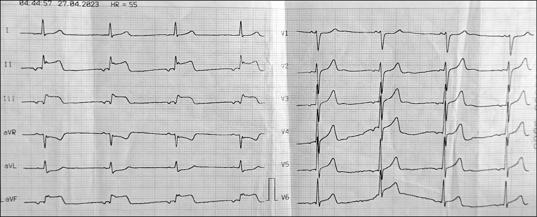
Myocarditis is often difficult to diagnose. The diagnostic difficulties include nonspecific symptoms or a “vague” clinical picture, absence of pathognomonic signs during physical examination, and endomyocardial biopsy, which is the “gold standard” of diagnosis of myocarditis, being an invasive procedure that is performed under strict indications in certain patients. Nevertheless, as radiology is rapidly developing, clinicians are now able to noninvasively diagnose symptoms of inflammatory myocardial damage, including edema and myocardial fibrosis, using cardiac magnetic resonance imaging. This article presents the clinical case of a young patient with symptoms of acute coronary syndrome, who showed no evidence of coronary artery disease. Myocarditis was suspected because of increased activity of cardiospecific enzymes and high levels of inflammatory markers, pronounced electrocardiography changes with positive dynamics, and recent infection. Magnetic resonance imaging was used to confirm myocarditis diagnosis. Thus, this case study demonstrates the role of imaging techniques in the differential diagnosis of ischemic and inflammatory heart diseases.
Full Text
##article.viewOnOriginalSite##About the authors
Natalia G. Poteshkina
The Russian National Research Medical University named after N.I. Pirogov; Moscow City Hospital 52
Author for correspondence.
Email: nat-pa@yandex.ru
ORCID iD: 0000-0001-9803-2139
SPIN-code: 2863-4840
MD, Dr. Sci. (Med.), Professor
Russian Federation, Moscow; MoscowElena A. Kovalevskaya
The Russian National Research Medical University named after N.I. Pirogov; Moscow City Hospital 52
Email: tolyaaa@mail.ru
ORCID iD: 0000-0002-0787-4347
SPIN-code: 8853-2700
MD, Cand. Sci. (Med.), Assistant professor
Russian Federation, Moscow; MoscowValentin E. Sinitsyn
Lomonosov Moscow State University Medical Research and Educational Center
Email: vsini@mail.ru
ORCID iD: 0000-0002-5649-2193
SPIN-code: 8449-6590
MD, Dr. Sci. (Med.), Professor
Russian Federation, MoscowElena A. Mershina
Lomonosov Moscow State University Medical Research and Educational Center
Email: elena_mershina@mail.ru
ORCID iD: 0000-0002-1266-4926
SPIN-code: 6897-9641
MD, Cand. Sci. (Med.), Assistant professor
Russian Federation, MoscowDaria A. Filatova
Lomonosov Moscow State University Medical Research and Educational Center
Email: dariafilatova.msu@mail.ru
ORCID iD: 0000-0002-0894-1994
SPIN-code: 2665-5973
Russian Federation, Moscow
Galina B. Selivanova
The Russian National Research Medical University named after N.I. Pirogov
Email: galina.selivanova@rambler.ru
ORCID iD: 0000-0003-2980-9754
SPIN-code: 9711-5041
MD, Dr. Sci. (Med.), Professor
Russian Federation, MoscowYavilika R. Shashkina
Moscow City Hospital 52
Email: yavilika-medik@mail.ru
ORCID iD: 0000-0002-2194-0785
Russian Federation, Moscow
References
- Ammirati E, Moslehi JJ. Diagnosis and Treatment of Acute Myocarditis: A Review. JAMA. 2023;329(13):1098–1113. doi: 10.1001/jama.2023.3371
- Caforio ALP, Calabrese F, Angelini A, et al. A prospective study of biopsy-proven myocarditis: prognostic relevance of clinical and aetiopathogenetic features at diagnosis. European Heart Journal. 2007;28(11):1326–1333. doi: 10.1093/eurheartj/ehm076
- Leone O, Veinot JP, Angelini A, et al. 2011 Consensus statement on endomyocardial biopsy from the Association for European Cardiovascular Pathology and the Society for Cardiovascular Pathology. Cardiovascular Pathology. 2012:21(4):245–274. doi: 10.1016/j.carpath.2011.10.001
- Arutyunov GB, Paleev FN, Moiseeva OM, et al. 2020 Clinical practice guidelines for Myocarditis in adults. Russian Journal of Cardiology. 2021;26(11):4790. (In Russ) doi: 10.15829/1560-4071-2021-4790
- Schultz JC, Hilliard AA, Cooper LT, et al. Diagnosis and Treatment of Viral Myocarditis. Mayo Clinic Proceedings. 2009;84(11):1001–1009. doi: 10.1016/s0025-6196(11)60670-8
- Caforio ALP, Pankuweit S, Arbustini E, et al. Current state of knowledge on aetiology, diagnosis, management, and therapy of myocarditis: a position statement of the European Society of Cardiology Working Group on Myocardial and Pericardial Diseases. European Heart Journal. 2013;34(33):2636–2648. doi: 10.1093/eurheartj/eht210
- Friedrich MG, Sechtem U, Schulz-Menger J, et al. Cardiovascular Magnetic Resonance in Myocarditis: A JACC White Paper. Journal of the American College of Cardiology. 2009;53(17):1475–1487. doi: 10.1016/j.jacc.2009.02.007
- Tijmes FS, Thavendiranathan P, Udell JA, et al. Cardiac MRI Assessment of Nonischemic Myocardial Inflammation: State of the Art Review and Update on Myocarditis Associated with COVID-19 Vaccination. Radiology: Cardiothoracic Imaging. 2021;3(6):e210252. doi: 10.1148/ryct.210252
- Srichai MB, Lim RP, Lath N, et al. Diagnostic performance of dark-blood T2-weighted CMR for evaluation of acute myocardial injury. Investigative Radiology. 2013;48(1):24–31. doi: 10.1097/rli.0b013e3182718672
- Galán-Arriola C, Lobo M, Vílchez-Tschischke JP, et al. Serial Magnetic Resonance Imaging to Identify Early Stages of Anthracycline-Induced Cardiotoxicity. Journal of the American College of Cardiology. 2019;73(7):779–791. doi: 10.1016/j.jacc.2018.11.046
- Blagova OV, Pavlenko EV, Varionchik NV, et al. Myocarditis as a legitimate phenomenon in patients with primary noncompaction myocardium: diagnosis, treatment and impact on outcomes. Russian Journal of Cardiology. 2018;23(2):44–52. (In Russ) doi: 10.15829/1560-4071-2018-2-44-52
- Filatova DA, Mershina EA, Sinitsyn VE. COVID-19-related cardiac lesion: The questions of pathogenesis and diagnostics. Digital Diagnostics. 2023;4(2):156−169. (In Russ) doi: 10.17816/DD284706
Supplementary files


