


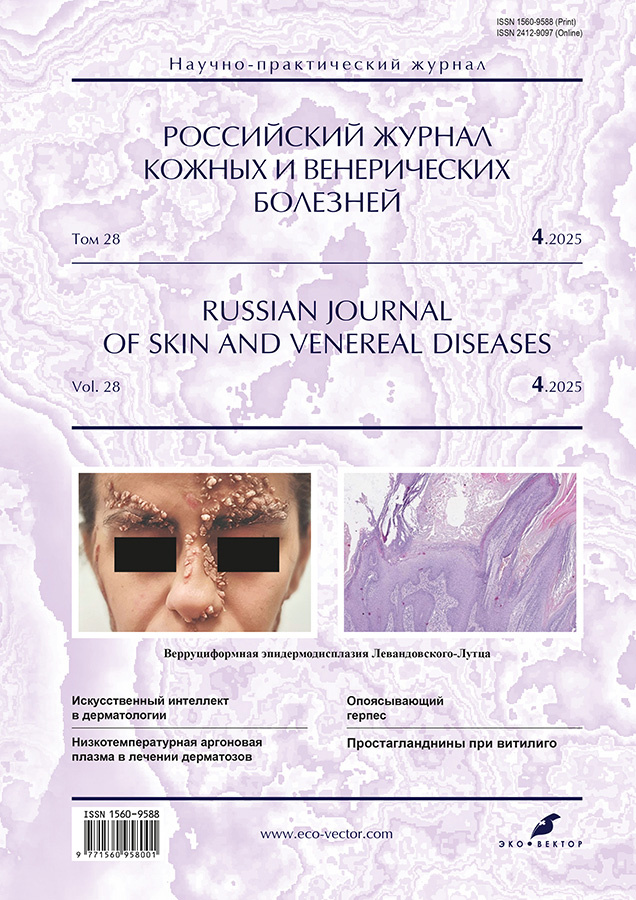
Lewandowsky–Lutz verruciform epidermodysplasia: a brief review and case report
- Authors: Belysheva T.S.1, Zelenova E.E.1,2, Prokofyev A.A.3, Semenova V.V.1,2, Sparber P.A.4, Kletskaya I.S.5, Aleskerova G.A.6, Valiev T.T.1, Nasedkina T.V.2
-
Affiliations:
- N.N. Blokhin National Medical Research Center of Oncology
- Engelhardt Institute of Molecular Biology
- Dermatology Center Petrovka 15
- Medical Genetic Research Center named after academician N.P. Bochkov
- Russian National Research Medical University named after N.I. Pirogov
- National Center of Oncology
- Issue: Vol 28, No 4 (2025)
- Pages: 369-380
- Section: DERMATO-ONCOLOGY
- URL: https://bakhtiniada.ru/1560-9588/article/view/350465
- DOI: https://doi.org/10.17816/dv679046
- EDN: https://elibrary.ru/VBUXDQ
- ID: 350465
Cite item

Abstract
Verruciform (warty) epidermodysplasia (Lewandowsky–Lutz syndrome) is a rare autosomal recessive disease manifested by multiple cutaneous lesions associated with human papillomavirus infection. To date, no more than 500 patients with this form of genodermatosis have been reported worldwide. Hereditary forms of verruciform (warty) epidermodysplasia are caused by inactivating mutations in the TMC6, TMC8, CIB1, RHOH, and IL7 genes in a homozygous or compound heterozygous state. A non-classical variant of hereditary verruciform (warty) epidermodysplasia has also been described; it is associated with primary T-cell immunodeficiency resulting from mutations in the CORO1A, CARMIL2, DCLRE1C, DOCK8, ECM1, GATA2, LCK, MST1, RASGRP1, and TPP2 genes. Acquired verruciform (warty) epidermodysplasia usually develops in patients with secondary immunodeficiency of various origins.
The low prevalence and insufficient clinical diagnostic experience explain the lack of standardized therapy protocols and clinical guidelines for verruciform (warty) epidermodysplasia. Several researchers and clinicians have reported treatment of this condition using chemotherapeutic agents, immunomodulators, interferons, retinoids, and other drugs in combination with surgical excision or physical methods of lesion destruction.
This article presents a case report of aggressive verruciform (warty) epidermodysplasia without preceding immunodeficiency or alterations in key genes, describing the experience of treating the patient with a retinoid in combination with CO2 laser destruction of cutaneous lesions.
Full Text
##article.viewOnOriginalSite##About the authors
Tatiana S. Belysheva
N.N. Blokhin National Medical Research Center of Oncology
Email: klinderma@bk.ru
ORCID iD: 0000-0001-5911-553X
SPIN-code: 2645-4049
MD, Dr. Sci. (Medicine)
Russian Federation, 23 Kashirskoe sh, Moscow, 115478Ekaterina E. Zelenova
N.N. Blokhin National Medical Research Center of Oncology; Engelhardt Institute of Molecular Biology
Author for correspondence.
Email: zelenovayeye@gmail.com
ORCID iD: 0000-0002-2197-8863
SPIN-code: 6823-6353
Russian Federation, 23 Kashirskoe sh, Moscow, 115478; Moscow
Alexander A. Prokofyev
Dermatology Center Petrovka 15
Email: alex-prok3@mail.ru
ORCID iD: 0000-0002-2466-785X
SPIN-code: 7891-8285
Russian Federation, Moscow
Vera V. Semenova
N.N. Blokhin National Medical Research Center of Oncology; Engelhardt Institute of Molecular Biology
Email: sulpiridum@yandex.ru
ORCID iD: 0000-0002-9705-1001
SPIN-code: 9014-2847
Russian Federation, 23 Kashirskoe sh, Moscow, 115478; Moscow
Peter A. Sparber
Medical Genetic Research Center named after academician N.P. Bochkov
Email: psparber93@gmail.com
ORCID iD: 0000-0002-9160-0794
SPIN-code: 3879-2993
MD, Cand. Sci. (Medicine)
Russian Federation, MoscowIrina S. Kletskaya
Russian National Research Medical University named after N.I. Pirogov
Email: ikletskaya@gmail.com
ORCID iD: 0000-0002-8552-7682
SPIN-code: 1046-3870
Russian Children's Clinical Hospital
Russian Federation, MoscowGunel A. Aleskerova
National Center of Oncology
Email: aleskerova@rambler.ru
ORCID iD: 0000-0001-7514-5413
MD, Cand. Sci. (Medicine)
Azerbaijan, BakuTimur T. Valiev
N.N. Blokhin National Medical Research Center of Oncology
Email: timurvaliev@mail.ru
ORCID iD: 0000-0002-1469-2365
SPIN-code: 9802-8610
MD, Dr. Sci. (Medicine)
Russian Federation, 23 Kashirskoe sh, Moscow, 115478Tatiana V. Nasedkina
Engelhardt Institute of Molecular Biology
Email: tanased06@rambler.ru
ORCID iD: 0000-0002-2642-4202
SPIN-code: 3741-8214
Dr. Sci. (Biology)
Russian Federation, MoscowReferences
- Belysheva TS, Nasedkina TV, Semenova VV, et al. Cancer-associated genodermatoses. Russian Journal of Pediatric Hematology and Oncology. 2022;9(2):60–74. doi: 10.21682/2311-1267-2022-9-2-60-74 EDN: IMMNFF
- Lewandowsky F, Lutz W. Ein fall einer bisher nicht beschriebenen hauterkrankung (epidermodysplasia verruciformis). Archiv für Dermatologie und Syphilis. 1922;141(2):193–203. doi: 10.1007/bf01938833
- Imahorn E, Yüksel Z, Spoerri I, et al. Novel TMC8 splice site mutation in epidermodysplasia verruciformis and review of HPV infections in patients with the disease. J Eur Acad Dermatol Venereol. 2017;31(10):1722–1726. doi: 10.1111/jdv.14431
- Vohra S, Sharma NL, Shanker V, et al. Autosomal dominant epidermodysplasia verruciformis: A clinicotherapeutic experience in two cases. Indian J Dermatol Venereol Leprol. 2010;76(5):557–561. doi: 10.4103/0378-6323.69092
- Burger B, Itin PH. Epidermodysplasia verruciformis. Curr Probl Dermatol. 2014;45:123–131. doi: 10.1159/000356068
- Oliveira WR, Rady PL, Festa C, et al. Skin cancer in epidermodysplasia verruciformis patients from Brazil. J Eur Acad Dermatol Venereol. 2006;20(9):1154–1156. doi: 10.1111/j.1468-3083.2006.01654.x
- Patel T, Morrison LK, Rady P, Tyring S. Epidermodysplasia verruciformis and susceptibility to HPV. Dis Markers. 2010;29(3-4):199–206. doi: 10.3233/DMA-2010-0733
- Howley PM, Pfister HJ. Beta genus papillomaviruses and skin cancer. Virology. 2015;(479-480):290–296. doi: 10.1016/j.virol.2015.02.004
- Edwards L, Reutter JC, Foster TE, et al. Perianal epidermodysplasia verruciformis associated with human papillomavirus 5 after a renal transplant. J Low Genit Tract Dis. 2017;21(3):e35–e36. doi: 10.1097/LGT.0000000000000316
- Rogers HD, Macgregor JL, Nord KM, et al. Acquired epidermodysplasia verruciformis. J Am Acad Dermatol. 2009;60(2):315–320. doi: 10.1016/j.jaad.2008.08.035
- Bostan E, Akdogan N, Gokoz O. Epidermodysplasia verruciformis after hematopoietic stem cell transplantation in a patient with severe combined immunodeficiency syndrome. Am J Dermatopathol. 2021;43(5):e65–e67. doi: 10.1097/DAD.0000000000001918
- Myers DJ, Kwan E, Fillman EP. Epidermodysplasia verruciformis [2023 Jan 29]. In: StatPearls [Internet]. Treasure Island (FL): StatPearls Publishing; 2023.
- Orth G. Genetics of epidermodysplasia verruciformis: Insights into host defense against papillomaviruses. Semin Immunol. 2006;18(6):362–374. doi: 10.1016/j.smim.2006.07.008
- De Jong SJ, Imahorn E, Itin P, et al. Epidermodysplasia verruciformis: Inborn errors of immunity to human beta-papillomaviruses. Front Microbiol. 2018;9:1222. doi: 10.3389/fmicb.2018.01222
- Nindl I, Gottschling M, Stockfleth E. Human papillomaviruses and non-melanoma skin cancer: Basic virology and clinical manifestations. Dis Markers. 2007;23(4):247–259. doi: 10.1155/2007/942650
- Ul Bari A, Yasmin R, Ahmed A. Epidermodysplasia verruciformis: A rare genodermatosis with risk of malignant transformation. J Pakistan Association Dermatol. 2006;16(4):242–245.
- Olczak P, Wong M, Tsai HL, et al. Vaccination with human alphapapillomavirus-derived L2 multimer protects against human betapapillomavirus challenge, including in epidermodysplasia verruciformis model mice. Virology. 2022;575:63–73. doi: 10.1016/j.virol.2022.08.006
- Montero-Vilchez T, Martinez-Lopez A, Rodriguez-Tejero A, et al. Epidermodysplasia verruciformis and breast cancer: Casual or causal? Indian J Dermatol. 2022;67(1):94. doi: 10.4103/ijd.ijd_830_20
- Zhang B, Xing H, Rui H, et al. Epidermodysplasia verruciformis mimicking pityriasis versicolor. Pediatr Investig. 2021;5(4):325–326. doi: 10.1002/ped4.12288
- Kirchhof MG, Au S. Brazilian waxing and human papillomavirus: A case of acquired epidermodysplasia verruciformis. CMAJ. 2015;187(2):126–128. doi: 10.1503/cmaj.140198
- Rajabi MT, Ghasemi H, Safizadeh M, et al. Conjunctival squamous cell carcinoma with intraocular invasion after radiotherapy in epidermodysplasia verruciformis. Can J Ophthalmol. 2014;49(2):e43–46. doi: 10.1016/j.jcjo.2013.12.009
- Alshammari R, Al-Issa A, Ghobara YA. Epidermodysplasia verruciformis: A rare case report. Cureus. 2020;12(7):e9046. doi: 10.7759/cureus.9046
- Yoshida R, Kato T, Kawase M, et al. Two sisters reveal autosomal recessive inheritance of epidermodysplasia verruciformis: A case report. BMC Dermatol. 2014;14:12. doi: 10.1186/1471-5945-14-12
- Sharma S, Barman KD, Sarkar R, et al. Efficacy of oral zinc therapy in epidermodysplasia verruciformis with squamous cell carcinoma. Indian Dermatol Online J. 2014;5(1):55. doi: 10.4103/2229-5178.126034
- Hoffner MV, Camacho FM. Surgical treatment of epidermodysplasia verruciformis. Dermatol Surg. 2010;36(3):363–367. doi: 10.1111/j.1524-4725.2009.01446.x
- Fang F, Zhao L, Jiang MJ, et al. Epidermodysplasia verruciformis with severe hand and foot deformity successfully treated with surgical excision. J Plast Reconstr Aesthet Surg. 2008;61(3):338–341. doi: 10.1016/j.bjps.2006.01.013
- Ginarte M, Peteiro C, Toribio J. Keloid formation induced by isotretinoin therapy. Int J Dermatol. 1999;38(3):228–229. doi: 10.1046/j.1365-4362.1999.00597.x
- Dogan G. Possible isotretinoin-induced keloids in a patient with Behçet’s disease. Clin Exp Dermatol. 2006;31(4):535–537. doi: 10.1111/j.1365-2230.2006.02140.x
- Mahadevappa OH, Mysore V, Viswanath V, et al. Surgical outcome in patients taking concomitant or recent intake of oral isotretinoin: A multicentric study-ISO-AIMS study. J Cutan Aesthet Surg. 2016;9(2):106–114. doi: 10.4103/0974-2077.184054
- Guadanhim LR, Gonçalves RG, Bagatin E. Observational retrospective study evaluating the effects of oral isotretinoin in keloids and hypertrophic scars. Int J Dermatol. 2016;55(11):1255–1258. doi: 10.1111/ijd.13317
Supplementary files


